

Veterinary paediatric dermatology is a field that calls for diagnostic rigour and therapeutic caution. Puppies and kittens, whose immune systems and skin are still maturing, present clinical pictures that differ markedly from those seen in adults. At the last ESVD congress in Bilbao, our colleague Ana Oliveira reviewed in detail the specific features of paediatric dermatology in puppies and kittens, both diagnostic and therapeutic.
These young patients present a remarkably wide range of skin diseases: fungal or bacterial infections, parasitic infestations, immune-mediated dermatoses and congenital connective tissue disorders. Each of these entities calls for an appropriate diagnostic approach and reasoned treatment choices that take account of the constraints of the animal’s age, weight and breed.
The initial history: the essential questions
The animal’s origin and the context in which the lesions appeared
Every paediatric dermatology consultation starts with a rigorous history, some elements of which are of prime diagnostic value. The animal’s origin is the very first piece of information to obtain: a puppy or kitten from a breeder is not exposed to the same epidemiological risks as an animal adopted from a shelter. In group settings, animal density and hygiene conditions favour the transmission of many pathogens, whether fungal, bacterial or parasitic.
The age at which the first clinical signs appeared is also central. In practice, owners often consult several weeks or even months after the problem began, which sometimes complicates interpretation of the lesions observed and can mislead as to the true timeline of the disease. The clinician must always try to reconstruct this timeline precisely, with targeted questions about the first signs observed, where they started and how they progressed over time. This reconstruction is all the more important because some paediatric conditions, notably dermatophytosis and parasitic infestations, can progress silently for several weeks before the lesions become clinically obvious.
Involvement of littermates
The question of whether other animals in the litter are affected is particularly important in parasitic and infectious conditions. Simultaneous involvement of several animals from the same litter points strongly to a contagious cause and should lead to examination of all potentially exposed individuals, whether other pets or, in some cases, the people in the household. This epidemiological information is crucial in guiding not only the diagnosis but also the extent of the therapeutic and preventive measures to be put in place. In some conditions with high zoonotic potential, such as dermatophytosis due to Microsporum canis, informing owners about the risk of transmission to humans is an integral part of management.
Dermatophytosis in young patients
Clinical presentation and variability of lesions
Dermatophytosis is one of the most common infectious diseases in puppies and kittens. Its clinical presentation, however, is disconcertingly variable, which calls for constant diagnostic vigilance. The classically described lesion, a circular area of alopecia with variable scaling and no notable pruritus, is certainly the most typical picture, but it is far from the rule. As a clinical example, a three-month-old French Bulldog may present with circular areas of alopecia, variable scaling and no pruritus, on the shoulder, armpit and head. Faced with this picture, the differential diagnosis includes dermatophytosis, bacterial folliculitis and demodicosis, which justifies a basic work-up from the outset.
In kittens, and particularly in long-haired breeds such as the Persian, scaling may be the only sign, with no obvious alopecia. In these situations, the condition risks being wrongly dismissed by the owner, or even by an inexperienced veterinarian, who puts the scaly coat down to poor grooming or an unsuitable diet. Generalised scaling in a young cat may, however, indicate diffuse dermatophytosis, whose zoonotic and contagious potential is real: the animal may transmit the infection to other animals in the household and to people in close contact with it.
In dogs, atypical presentations also exist, notably in the form of a kerion, an oozing nodular lesion that histologically corresponds to a purulent neutrophilic infiltrate with macrophages and fungal hyphae, or of pustular and crusting dermatitis. Faced with pustular crusting dermatitis that does not respond to antibiotics, the possibility of dermatophytosis caused by a species other than Microsporum canis must always be considered. This vigilance is all the more necessary because other, less common dermatophyte species can produce misleading clinical pictures, and their identification requires a full fungal culture with morphological examination of the macroconidia, as well as fungal histological stains.
Diagnostic tools: from the Wood’s lamp to fungal culture
The basic work-up for any suspicious paediatric alopecia includes trichography, deep skin scraping and Wood’s lamp examination. The latter has a special place in the assessment of young patients: it is non-invasive and quickly reveals the green fluorescence characteristic of Microsporum canis on affected hair shafts. It is essential to examine the whole body surface, not just the clinically visible areas of alopecia. Fluorescent hairs may be present in areas that look grossly normal, such as around the eyes, the edges of the pinnae and the muzzle, revealing more extensive involvement than clinical examination alone suggests. The fluorescence seen with the Wood’s lamp is a bright green that specifically affects the hair shafts and not scale or crusts, an important interpretive criterion for not confusing true dermatophyte fluorescence with light artefacts.
Fluorescent hairs have a dual practical value: sampling them specifically improves the quality of trichography, in which arthrospores arranged in a sheath around the hair shaft confirm the infectious nature of the lesion and rule out accidental environmental contamination. These same hairs are also the best material for inoculating a fungal culture. Using fluorescent hairs for trichography and culture therefore offers a considerable diagnostic advantage over blind sampling, significantly increasing the likelihood of demonstrating the pathogen.
In clinical practice, DTM (Dermatophyte Test Medium) is commonly used: a colour change of the medium from yellow to red, together with the growth of whitish colonies, is a warning sign that justifies further analysis. This involves morphological identification of the macroconidia, the only way to confirm that the organism is a dermatophyte and to determine the species responsible. In animals with generalised involvement, brushing the whole coat with a new toothbrush before inoculating the culture medium is a simple and effective technique, particularly suited to diffuse cases in which there are too many lesions for systematic localised sampling.
Age-appropriate treatment strategies
Treatment of paediatric dermatophytosis combines topical measures and, where needed, systemic treatment. Clipping the coat, long recommended, is now approached with caution because of the risk of spreading fungal spores during the procedure. Stepping up environmental hygiene, on the other hand, remains essential, with regular decontamination of surfaces, bedding and items in contact with the animal.
Topically, lime sulphur dips have been used for a long time and are recognised for their efficacy and satisfactory safety profile. Since their availability varies from country to country, 0.2 % enilconazole or a shampoo combining chlorhexidine and miconazole are valid alternatives widely available in most European countries.
When systemic treatment is needed, particularly in generalised forms or to prevent transmission to littermates and owners, itraconazole can be prescribed in kittens weighing more than one kilogram or older than six weeks. In puppies its use is off-label, which means informing the owner and documenting the clinical decision in the animal’s record. This transparency is essential to build a relationship of trust with the owner and to comply with the regulatory obligations governing off-label prescribing in veterinary medicine.
Bacterial skin infections: folliculitis and impetigo
Clinical distinctions and diagnostic implications
Superficial bacterial infections are another common reason for dermatology consultations in young dogs and cats. The distinction between bacterial folliculitis and impetigo has clinical importance that goes beyond classification, because it directly determines the search for an underlying cause.
Bacterial folliculitis is an infection located within the hair follicle. The resulting pustules are small and pierced by a hair at their centre, a pathognomonic sign when present. Circular lesions with peripheral erythema and central hyperpigmentation are also seen, reflecting a course of some duration. Lesions are mainly distributed over the abdomen and the medial thighs. This presentation is sometimes discovered by chance, for example at a pre-operative examination: a young bitch prepared for spaying may reveal, after clipping, numerous papules and pustules on the belly and the inner hind legs, with here and there a typical pustule with a hair emerging exactly from its centre. In this context an underlying cause must always be sought: demodicosis, food or environmental hypersensitivity may provide the ground that favours bacterial proliferation.
Impetigo is distinguished by the intraepidermal location of the infection, which produces larger pustules. Unlike folliculitis, impetigo generally occurs without an identifiable underlying cause and mainly affects healthy puppies. Cytology of both entities reveals a neutrophilic infiltrate with cocci, intracellular or extracellular depending on the degree of phagocytic activity. This cytological similarity underlines the importance of clinical assessment in distinguishing the two, notably by observing the size of the pustules and their relationship to the hair follicle.
Treatment approach
Impetigo can be treated topically alone: local application of chlorhexidine is generally enough to resolve the infection without systemic antibiotics. For bacterial folliculitis, topical treatment is also sufficient in most cases, provided the predisposing cause is identified and corrected. Systemic antibiotics should be reserved for situations in which topical treatment alone is insufficient or impractical, to limit the risk of selecting bacterial resistance, a problem of growing importance in veterinary and human medicine alike.
Juvenile cellulitis: a dermatosis of immune origin
Clinical picture and diagnostic criteria
Juvenile cellulitis is a skin disease characteristic of puppies aged roughly three weeks to four months. The clinical picture typically begins with acute facial swelling, mainly around the eyes and nose, quickly followed by pustules and more or less marked exudation. Skin lesions may also appear in the perianal region, although this location is less consistent and facial involvement remains the main guiding sign.
The most characteristic clinical sign, and probably the most useful for the diagnosis, is enlargement of the mandibular lymph nodes, which can be spectacular and out of proportion to the size of the animal. This sign, together with the predominantly facial location and the age group concerned, should alert the clinician and point immediately to this diagnosis.
On cytology, in the absence of secondary bacterial infection, the lesions contain a sterile pyogranulomatous exudate composed of neutrophils and macrophages without bacteria. This inflammatory profile points to immune dysregulation, the most commonly proposed but not yet definitively established aetiopathogenic hypothesis. A genetic predisposition has been suggested in view of the breed distribution of the disease and the sometimes simultaneous involvement of several individuals from the same litter, an additional epidemiological argument for a hereditary component.
Treatment
The response to immunomodulatory treatment is generally rapid and is in itself a retrospective diagnostic argument. Prednisolone given for two to four weeks is the reference protocol. Topical antiseptic treatment with chlorhexidine is always added to prevent secondary bacterial infections, which could complicate the course and leave scars that permanently affect the animal’s appearance. Preventing these scars is indeed one of the main reasons to act quickly as soon as the diagnosis is made.
Ectoparasitic diseases: Cheyletiella, sarcoptic mange and demodicosis
Cheyletiellosis
Cheyletiella spp. is a surface mite that causes diffuse scaling, often with pruritus of varying intensity. These mites, recognised by their claw-like hooks and visible under the light microscope, can be demonstrated by superficial scraping or tape strips. Direct microscopy readily identifies their distinctive morphology.
Sarcoptic mange
Sarcoptic mange causes intense, often severe pruritus, with skin excoriations and a positive pinnal-pedal reflex on clinical examination. The ear margins are classically affected, with characteristic papular and crusting lesions. Cytology of these papules typically shows eosinophils, reflecting a hypersensitivity reaction to the mite. Superficial skin scraping may identify Sarcoptes scabiei mites, although their numbers are sometimes low and the sensitivity of this test is limited, so a negative result is not enough to rule out the diagnosis.
The isoxazolines, whose marketing authorisation now includes the treatment of sarcoptic mange, have greatly simplified the management of this disease. Their high efficacy and satisfactory tolerability make them the treatment of choice in young patients, offering a much more convenient alternative to the old protocols based on amitraz or injectable ivermectin, which involved more constraints and potential risks in young animals.
Juvenile demodicosis
Juvenile demodicosis occurs in two distinct clinical forms whose prognostic significance differs markedly. The localised form, with a few focal areas of alopecia, mainly around the eyes and nose or on the distal limbs, generally resolves spontaneously and does not always require specific treatment. The generalised form, by contrast, is a serious disease that can affect the whole body surface, with extensive alopecia, hyperkeratosis, hyperpigmentation and frequently an associated superficial bacterial infection, shown by pustules and a characteristic bluish hyperpigmentation, clearly visible on the abdominal skin of lightly pigmented animals.
As an example, a French Bulldog acquired at two months of age, with some initial hair loss that went unnoticed, may progress to severe generalised demodicosis affecting the whole body by the time of consultation, sometimes several months after the disease began. The clinical picture is then striking: almost no hair over large areas, marked hyperkeratosis, diffuse hyperpigmentation and superficial bacterial folliculitis on the belly, with the characteristic pustules and the typical bluish hyperpigmentation that accompanies pyoderma associated with chronic demodicosis.
Diagnosis relies on deep skin scraping, which reveals large numbers of Demodex mites in the hair follicles. In hard-to-reach areas, notably around the eyes, trichography, performed by careful manual plucking of hairs, offers a practical and effective alternative. Applying adhesive tape to squeezed skin is a third approach, particularly useful in uncooperative patients or in areas where scraping is technically difficult.
The arrival of the isoxazolines has profoundly transformed the management of generalised demodicosis. Before they were available, severe forms of this disease were real therapeutic challenges, particularly in puppies of brachycephalic breeds. Today, complete clinical remission, confirmed by two consecutive negative skin scrapings two weeks apart, is regularly achieved with these drugs. This cure criterion, combining complete clinical remission with two consecutive negative scrapings, must be strictly respected before stopping treatment is considered, to avoid relapse. Regular monitoring of patients on treatment nevertheless remains essential, and the frequency of rechecks should be adapted to the initial severity and the clinical course of each individual.
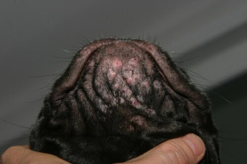
Juvenile demodicosis
Skin hypersensitivities in puppies
Adverse food reaction: diagnostic approach and elimination protocol
Once infectious and parasitic causes have been ruled out, persistent pruritus in a puppy should lead to consideration of a hypersensitivity. Adverse food reaction can appear very early, from four months of age, with pruritus mainly affecting the abdomen, the interdigital spaces and the axillae. Erythema, excoriations and, when the course is prolonged, progressive hyperpigmentation are the usual skin signs. A puppy may thus be presented with abdominal pruritus that began at four months of age, with excoriations, diffuse erythema, some hyperpigmentation reflecting the duration of the problem, and marked interdigital erythema.
The elimination diet is the reference diagnostic test. In a puppy, it must take account of the animal’s age, weight and breed, in order to choose a diet suited to the specific nutritional needs of a growing body. A six-month-old Yorkshire Terrier will reach adult size relatively quickly, which widens the dietary options available, whereas a Great Dane of the same age will keep growing until about eighteen months, which imposes stricter nutritional constraints. Manufacturers of veterinary diets based on hydrolysed or novel proteins can be consulted to confirm that the intended product meets the animal’s needs. If there is any doubt about the nutritional adequacy of a home-made diet based on a novel protein, the advice of a veterinary nutritionist should be sought to ensure the diet is balanced and to prevent deficiencies that could compromise the young animal’s development.
The recommended minimum duration of the elimination diet is eight weeks on average, bearing in mind that some patients may need ten to twelve weeks to show significant clinical improvement. If there is improvement, partial or complete, a provocation test by reintroducing the original diet is needed to confirm the diagnosis. If this test reproduces the clinical signs, the diagnosis of adverse food reaction is confirmed. If there is no improvement, or if improvement remains partial after a correctly conducted diet, atopic dermatitis should be considered.
Aeroallergen-induced atopic dermatitis: an entity diagnosed ever earlier
Aeroallergen-induced atopic dermatitis is a complex, multifactorial inflammatory skin disease resulting from the interaction between epidermal barrier abnormalities, an aberrant immune response to environmental allergens and a breed-related genetic predisposition. House dust mites, pollens and moulds are the most frequently involved allergen sources.
Clinical signs suggestive of atopic dermatitis can appear in the first months of life, particularly in strongly predisposed breeds such as the French Bulldog. Erythema of the axillary folds, interdigital spaces, periocular and perioral areas and abdomen is the classic picture. In puppies, owners sometimes interpret these behaviours, such as repeated paw licking and scratching of the belly, as normal play, which delays consultation. The insidious progression of the lesions eventually alerts attentive observers, as the erythema intensifies, scratch lesions become more numerous and the pruritic behaviour becomes a lasting part of the animal’s routine. A French Bulldog licking its paws and scratching its belly from eight months of age, with axillary, interdigital, periocular, perioral and abdominal erythema, is a representative example of this early clinical picture, which calls for a structured diagnostic approach without delay.
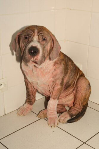
Atopic dermatitis can start very early in some breeds
Treatment of the atopic puppy: reactive phase and proactive strategy
Managing atopic dermatitis in puppies requires two complementary levels of intervention: reactive therapy to control pruritus quickly, and a proactive approach to maintain skin comfort in the long term.
Reactive therapy
Lokivetmab is the first-line systemic treatment in puppies, because it is authorised with no minimum age restriction. Given monthly by subcutaneous injection in Europe, it produces a response that varies between individuals, as in adults, and may be fully or partially effective depending on the case. This individual variability is a clinical reality to anticipate when informing the owner, to avoid disappointment and maintain treatment adherence even when the initial response is incomplete. In addition, prednisolone can be used for short periods, given once daily, to control severe pruritic flares. Caution is needed with a growing body, whose physical development and immune maturation could theoretically be affected by prolonged corticosteroid therapy, even though short courses are generally well tolerated. This calls for an open discussion with the owner, stressing that if the treatment is needed, it must be kept as short as possible.
Hydrocortisone aceponate applied topically once daily is an interesting alternative for localised pruritic areas such as the interdigital spaces or the abdomen. Its efficacy on circumscribed inflammatory foci is satisfactory, provided treatment is not prolonged excessively because of the risk of skin atrophy. Complementary topical treatments, such as spot-ons, mousses and chlorhexidine or moisturising wipes, are well tolerated by puppies and actively contribute to symptom control, particularly when baths cannot be given regularly. These formulations have the advantage of being easy for owners to use, which helps adherence when regular bathing is hard to maintain.
The primary goal of reactive therapy is to stop the pruritus and give the animal immediate comfort. It is possible, and often desirable, to combine several of these approaches to achieve this. A moisturising bath, combined if needed with a chlorhexidine shampoo when there is concurrent dysbiosis or superficial pyoderma, usefully complements drug treatment. When frequent bathing is not possible, spot-ons, mousses and wipes are practical alternatives that maintain a beneficial effect on the skin barrier between baths.
Proactive strategy
In the long term, and particularly in breeds with a strong atopic predisposition, a proactive strategy should be put in place as soon as inflammatory flares are under control. Supplementation with essential omega-3 and omega-6 fatty acids, or a diet naturally rich in these nutrients, helps maintain the integrity of the skin barrier and reduce background inflammation. Antihistamines, whose antipruritic efficacy is recognised as limited, can nevertheless be used in puppies as adjunctive treatment. Palmitoylethanolamide, a mast-cell inhibitor with mild antipruritic activity, can be introduced at any age; because its action is gradual, it belongs to long-term maintenance rather than crisis therapy, and introducing it early in the atopic puppy’s treatment may help reduce the intensity and frequency of inflammatory flares over time. Baths with a moisturising shampoo, combined if needed with a chlorhexidine shampoo when there is concurrent dysbiosis or superficial pyoderma, are a pillar of maintenance treatment, to be continued regularly even between flares.
Age restrictions for systemic treatments
The minimum age restrictions for the main immunomodulatory drugs available must be strictly respected: ciclosporin can be used from six months, oclacitinib from one year, immunocytokines from one year and allergen-specific immunotherapy from six months. Starting immunotherapy must be preceded by allergy testing, intradermal testing or serum IgE measurement, preferably carried out after one year of age. Testing too early risks failing to detect all the specific IgE in the serum, either because sensitisation is incomplete or because the puppy has not yet been exposed to all the seasonal allergens. Waiting until one year of age thus ensures full exposure to the different seasons and optimises the diagnostic value of the test, allowing an informed decision about immunotherapy based on allergy testing that truly reflects the animal’s sensitisation profile.
Skin diseases specific to kittens
Proliferative and necrotising otitis externa
Proliferative and necrotising otitis externa is a rare clinical entity described only in kittens, usually between two and six months of age. It presents as well-demarcated plaques covered with thick, adherent crusts, mainly at the entrance to the external ear canal, although facial lesions, with circular plaques on the lips, and scrotal lesions have been reported. The associated pruritus varies between individuals. The most common complication is secondary otitis, due to partial obstruction of the canal by the crusted plaques, which may cause ear pain and associated behavioural changes that owners describe as irritability or increased sensitivity when the head is touched.
The aetiopathogenesis of this disease remains poorly understood. Immunological arguments have, however, been put forward, notably the involvement of CD3 T lymphocytes in the inflammatory process, suggesting an aberrant, locally mediated immune response. Spontaneous resolution is possible in some patients as the immune system matures, which is consistent with the hypothesis of transient immune dysregulation linked to the immaturity of the lymphocyte system in young kittens.
Diagnosis rests on histopathology in diagnostically challenging cases, although in practice a typical clinical presentation in a kitten of the relevant age may justify starting treatment without prior histological confirmation. For treatment, topical tacrolimus and topical hydrocortisone aceponate, applied twice daily, are the first-line options. Oral steroids are rarely needed. Documented treatment responses are favourable, with complete clinical remission within two weeks of properly conducted topical treatment, as shown by cases in which kittens with both pinnae covered in thick plaques achieved complete remission after two weeks of twice-daily hydrocortisone aceponate.
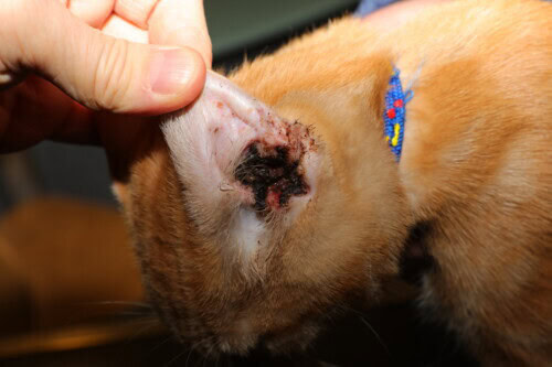
Proliferative and necrotising otitis externa in a kitten
Cutaneous asthenia: a congenital disease with no cure
Clinical and genetic features
Cutaneous asthenia, also known as veterinary Ehlers-Danlos syndrome, is a congenital connective tissue disease seen in dogs and cats and characterised by remarkable skin hyperextensibility. Owners describe the skin as tearing spontaneously at the slightest trauma, which reflects a profound mechanical fragility due to a structural abnormality of dermal collagen. The skin hyperextensibility is truly astonishing on clinical examination: owners can lift and stretch their animal’s skin to a degree far beyond what an examiner would expect. Paradoxically, these patients do not seem to show pain during tearing episodes, heal well and do not usually develop secondary infections, a remarkable feature of this disease.
The mode of transmission is hereditary, with autosomal dominant and recessive forms depending on the species and the mutations involved. Several genes have been identified in the different species affected. Neutering affected animals is imperative to prevent the mutation being passed on to offspring, and this recommendation must be clearly communicated to owners as soon as the diagnosis is made.
Long-term management
There is currently no curative treatment for cutaneous asthenia. Management is therefore purely symptomatic and relies on repeated suturing of skin wounds throughout the animal’s life. These procedures, which may be frequent depending on the individual and its activities, require regular veterinary follow-up and careful education of owners about the precautions needed to limit skin trauma. Restricting potentially traumatic activities, such as play with other animals or access to risky areas, and adapting the living environment are the pillars of preventing tears. Psychological support for owners is also important, because lifelong management of an animal with cutaneous asthenia can be a considerable emotional and financial burden, which should be anticipated from the moment the diagnosis is given.
Conclusion
Veterinary paediatric dermatology covers a broad spectrum of diseases, some directly linked to the immaturity of the immune system, others to infectious or parasitic agents that take advantage of the young body’s vulnerability, and others again to immune dysregulation or congenital connective tissue defects. Faced with a puppy or kitten with skin lesions, the diagnostic approach must be methodical and prioritised, starting with a precise history, including the animal’s origin, the age at which signs appeared and whether littermates are affected, before directing further tests according to the most likely hypotheses.
The diagnostic tools available, Wood’s lamp, trichography, skin scraping, cytology, fungal culture and histopathology, allow a reliable diagnosis when used in a targeted and reasoned way. In treatment, choices must take account of age, weight and breed, favouring drugs with a marketing authorisation suited to young patients and reserving powerful systemic treatments for situations in which the clinical benefit justifies the risk-benefit balance.
Recent advances with the isoxazolines for ectoparasitic diseases and lokivetmab for paediatric atopic dermatitis have profoundly changed practice, offering effective and better-tolerated treatment alternatives. Research questions remain open, notably on the aetiopathogenesis of juvenile cellulitis and of proliferative and necrotising otitis externa, whose underlying immune mechanisms are still partly unclear. Likewise, determining the optimal time to start specific immunotherapy in atopic puppies deserves thorough prospective studies, to define precise clinical and biological criteria to guide this decision.
Oliveira A. Dermatological diseases in paediatric patients. ESVD Conference, Bilbao, 2025
