

Geriatric dermatology is taking up a growing place in our daily practice, as a direct consequence of the longer life expectancy of dogs and cats. This longevity brings dermatoses that often reveal an underlying systemic disease, whose early recognition determines the prognosis. At the last ESVD congress, our colleague Ana Oliveira, who practises in Lisbon, presented the main ones.
The skin of the older dog and cat is no longer just a covering: it becomes the window through which an unsuspected visceral disease shows itself. Because of this link, managing the geriatric patient is not limited to symptomatic treatment of the skin lesion observed. It calls for a comprehensive diagnostic and therapeutic approach that takes account of the comorbidities common at this age, such as chronic kidney disease, degenerative heart disease or an endocrinopathy. Ageing of the skin is never just a cosmetic change: in a notable proportion of patients it comes with systemic and skin diseases whose diversity makes the differential diagnosis difficult.
It should be remembered that physiological skin ageing, regardless of any disease, leads to progressive thinning of the epidermis, reduced dermal blood supply and a decline in barrier function. These changes are not pathological in themselves, but they weaken the skin and make it more vulnerable to the expression of underlying dermatoses that would have remained silent in a younger animal.
1 Epitheliotropic cutaneous lymphoma
1.1 A striking epidemiological correlation
Ana Oliveira gives the example of an atopic dog of around ten years of age, whose pruritus had until then been well controlled on the usual protocol, and whose skin condition deteriorates: first moderate scaling appears, then the pruritus flares, and finally it becomes refractory to treatments that usually work. In this geriatric setting, suspicion of epitheliotropic cutaneous lymphoma must be the first differential diagnosis, even before considering a simple loss of control of the atopic dermatitis.
Epidemiological studies suggest that atopic dogs have an increased risk of developing this skin neoplasm.
This pattern is not unique to veterinary medicine. In human medicine, persistent atopic dermatitis is also thought to predispose to the late onset of epitheliotropic lymphoma, which strengthens the biological plausibility of the association seen in dogs. This parallel opens the way to translational research: the older atopic dog could be a useful spontaneous model for studying the mechanisms of lymphomatous transformation also described in humans.
1.2 Pathophysiological hypotheses: from chronic inflammation to lymphocyte clonality
The exact mechanism of this neoplastic transformation remains uncertain, and the scientific community has no certainty on this point. The most widely held hypothesis concerns the chronicity of atopic inflammation: uninterrupted antigenic stimulation, sustained by aeroallergens and by recurrent dysbiosis of the skin microbiota, could induce clonal hyperproliferation of T lymphocytes that are permanently activated in an inflammatory microenvironment. This hypothesis has not, however, been formally proven.
A second lead concerns a possible iatrogenic role of the immunomodulatory drugs used long term in atopic dermatitis. The key study on which this correlation rests included patients who had never been treated at the time of lymphoma diagnosis, while other animals were receiving certain anti-allergic treatments when the neoplasm was diagnosed. This temporal coincidence establishes no causal link, but it is worth bearing in mind, without calling into question the value of immunomodulatory treatments, whose benefit for the quality of life of the atopic patient remains real. The atopic background is itself a risk factor, regardless of the treatments given.
A third, less often mentioned hypothesis concerns chronic changes in the skin barrier. In the atopic dog, the constitutional abnormality of the epidermal barrier, together with reduced expression of certain structural proteins such as filaggrins, could promote increased and persistent antigen exposure of intraepidermal lymphocytes, creating over long periods a favourable ground for the emergence of an aberrant lymphocyte clone. This hypothesis remains speculative and would need to be confirmed by large longitudinal studies.
1.3 A varied clinical picture
The erythematous, scaling and exfoliative form
In an atopic patient whose disease is escaping control, two signals deserve particular attention. The first is the appearance of progressive alopecia on the trunk and limbs. The second concerns examination of the nasal planum, which is often telling: it frequently shows progressive leukoderma, together with loss of its usual cobblestone architecture.
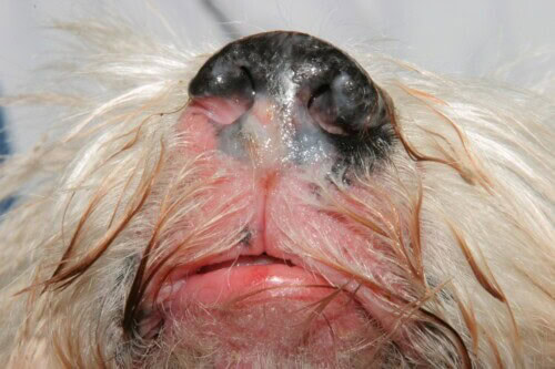
Leukoderma
Elsewhere on the skin, the neoplastic lymphocytic infiltrate causes orthokeratotic hyperkeratosis, which shows as broad, adherent scales together with erythroderma. This term refers to generalised erythema of frankly pathological intensity, to be distinguished from localised reactive erythema. A first case illustrated this triad: diffuse erythema, marked alopecia and leukonychia. This last sign deserves attention, because leukonychia should not be regarded as a simple physiological feature of old age, but should raise suspicion of lymphomatous infiltration of the claw structures. Other observations show similar changes of the nasal planum, with deep scaling close to severe exfoliative dermatitis, itself combined with erythema and extensive truncal alopecia.
In practice, these early signs are first interpreted as a worsening of the existing atopic dermatitis, which often explains a delay in diagnosis. Vigilance must therefore be at its highest whenever the clinical phenotype changes in an older atopic dog, even when the change at first seems minor.
Progression to plaques and nodules
Without treatment, the disease progresses. In proportion to the density of the lymphocytic infiltrate in the epidermis, the skin is remodelled and forms plaques, indurated and sharply demarcated from the neighbouring healthy skin. These plaques precede the appearance of nodules, at first a few millimetres in size, whose growth follows the aggressiveness of the neoplastic clone. The rate of growth varies from one individual to another, depending on variables such as the mitotic index observed on histopathology.
The tissue distension caused by nodular enlargement leads to ulceration of the stratum corneum. These epidermal breaches then provide a favourable substrate for opportunistic bacterial colonisation. Systematic cytology of the ulcer exudates is therefore required, to characterise and quantify any secondary septic component, which guides symptomatic management. The varied nature of this neoplasm also shows in single or multiple masses: an exophytic mass on a hind limb, for example, or an ulcerated mass destroying the metacarpal pad.
Mucosal and mucocutaneous forms
While exfoliative erythroderma and the nodular-ulcerative stage are the most marked forms of the disease, some presentations are limited to the oral mucosa or the mucocutaneous junctions. This topographical distinction is not trivial: it affects the prognosis, as we shall see below.
Oral involvement shows as ulcerative foci scattered over the oral mucosa, always surrounded by an erythematous halo reflecting local lymphocytic infiltration. The mucocutaneous variant of the nasal planum is marked by early depigmentation and loss of the cobblestone surface, followed by indolent ulcers that are slow to heal spontaneously. The disease often spreads to the philtrum and lip margins, which undergo the same changes. This form may also begin with simple bilateral, symmetrical depigmentation, later complicated by subtle lip erosions.
This very presentation led to the referral of a patient seen some years ago at her clinic, our colleague explains. Clinical examination showed focal crusted ulcers limited to two footpads in an older dog that was otherwise systemically well. In endemic areas of the Mediterranean basin, ulcerative-crusting pododermatitis first requires leishmaniosis to be ruled out. Epitheliotropic cutaneous lymphoma must nevertheless be included in the differential diagnosis from the earliest stages, or management will be delayed, and early management determines the prognosis.
This case is a reminder of the need for a systematic approach, not biased by regional assumptions: if the prevalence of leishmaniosis in some areas justifies considering it first, it should not lead to the premature exclusion of a rarer but equally serious differential diagnosis.
1.4 The diagnostic approach: from cytology to molecular biology
Cytology is the first step whenever epitheliotropic cutaneous lymphoma is suspected. It shows a proliferation of intermediate to large lymphocytes, with suggestive pleomorphism and nuclear atypia. Aberrant mitotic figures and the so-called “hand-mirror” morphology, characteristic of T lymphocytes, support the diagnosis at this early stage.
Confirmation rests on histopathology, performed on multiple skin biopsies taken from several sites to allow for the heterogeneity of the lesions. Histology shows the epitheliotropism of the neoplastic lymphocytes, which colonise the epidermis diffusely or in aggregates, the Pautrier microabscesses. This infiltration also reaches the follicular structures, which explains the alopecia and leukonychia seen clinically. The T phenotype of the neoplasm calls for immunohistochemical characterisation, which shows consistent positivity for CD3 and, frequently in dogs, for CD8, confirming the cytotoxic T-lymphocyte nature of the proliferation.
Putting together the history, clinical signs, cytology and histopathology makes the diagnosis possible in most cases. In the earliest macular stages, however, the histological architecture may lack specificity and make interpretation difficult. In these ambiguous cases, PCR analysis of lymphocyte clonality, known by the acronym PARR, provides a decisive argument: it distinguishes a neoplastic proliferation from a reactive inflammatory process by demonstrating uniform rearrangement of the antigen receptors, the signature of a monoclonal population. This test requires good-quality tissue or cytology samples; the quantity and quality of the extracted DNA determine the reliability of the result and the risk of false negatives, which is common in tissues fixed in formalin for a long time.
The diagnostic approach thus proceeds in steps: clinical suspicion based on the epidemiological context and the clinical signs, rapid cytological confirmation, then histopathological and immunohistochemical characterisation, supplemented if needed by molecular clonality analysis when uncertainty remains. This order optimises both the speed and the reliability of the diagnosis, two parameters that are decisive for subsequent management.
1.5 Prognostic factors
Median survival without treatment ranges from a few months to two years, with considerable variability depending on several clinical and histological factors. Forms limited to the oral mucosa or mucocutaneous junctions are associated with longer survival than diffuse skin involvement, which justifies precise topographical characterisation from the first examination. Likewise, a low mitotic index reflects indolent tumour growth and points to a more favourable prognosis.
1.6 Current treatment options
The conventional treatment relies mainly on sequential single-agent chemotherapy with lomustine, given at three-week intervals. The narrow therapeutic margin of this drug requires rigorous haematological and biochemical monitoring on the tenth day after each dose, to detect iatrogenic hepatotoxicity, neutropenia or thrombocytopenia, which may prevent the protocol from continuing. Complete remission is achieved in about thirty per cent of cases, although its duration cannot be predicted when treatment starts. Lomustine nevertheless prolongs survival compared with purely palliative care, which is worth explaining to owners.
Rabacfosadine currently offers an interesting alternative. Preliminary data show a reduction in clinical scores for erythema and alopecia with this treatment. The lack of any decrease in tumour volume in ulcerated masses, however, calls for further investigation before its use is extended to all presentations of the disease.
Whatever antitumour protocol is chosen, managing pruritus remains a constant challenge, because it impairs the animal’s daily well-being. It combines rigorous antipruritic treatment and, when secondary infections complicate the picture, targeted antibiotic therapy. Local skin hygiene, mild antiseptics and suitable emollients limit the discomfort of exudative and ulcerated lesions while making it easier to monitor their progress.
2 Superficial necrolytic dermatitis
Epitheliotropic lymphoma is not the only geriatric dermatosis in dogs. Superficial necrolytic dermatitis is also relatively common and mainly affects dogs approaching ten years of age. It is an example of systemic dysfunction in which liver changes silently precede the skin lesions, sometimes by several months.
2.1 Clinical presentation on the feet and face
Footpad hyperkeratosis often leads to fissuring and is frequently the first sign. It causes marked foot discomfort and lameness, often the reason for the first consultation. Typically several feet are affected, with involvement of all the pads, the interdigital spaces and the digits, together with erythema, crusted exudate and erosions of the nasal planum, the mucocutaneous junctions and bony prominences. The appearance of hyperkeratotic calluses on the elbows is a hepatic warning sign that calls for a full biochemical profile and an abdominal ultrasound without delay.
In advanced cases, foot discomfort is sometimes so severe that the animal is reluctant to walk, which may at first suggest an isolated orthopaedic problem, especially in older dogs in which osteoarthritis is common. This possible confusion justifies a systematic and careful examination of the feet in any geriatric patient with lameness of gradual onset, whatever the initial orthopaedic hypothesis.
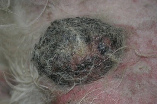
Superficial necrolytic dermatitis with testicular involvement
2.2 The hepatic and pancreatic metabolic origin
The metabolic basis of this condition lies in a fibrosing hepatopathy that causes profound catabolic hypoaminoacidaemia. This is often combined with concurrent diabetes mellitus or, in some presentations, a secreting glucagonoma, an endocrine pancreatic tumour whose secretion worsens the protein catabolism already compromised by liver failure. This dual hepatic and pancreatic origin explains the variety of clinical presentations and justifies a full systemic work-up as soon as the condition is suspected.
2.3 Combined histological and ultrasound diagnosis
The diagnosis of superficial necrolytic dermatitis rests on two complementary tests. On the one hand, skin histopathology shows a now well-known pattern, the so-called “French flag”, in reference to the three-coloured layering of the damaged epidermis: superficial hyperkeratosis, intercellular oedema in the middle layer and a hyperplastic basal layer. On the other, abdominal ultrasound reveals a dysplastic liver parenchyma, whose honeycomb appearance reflects regenerative hepatocyte nodules surrounded by bands of fibrosis and fatty tissue.
2.4 Palliative management
Treatment options remain limited. They are restricted to palliative correction of the nitrogen balance, with amino acid infusions and an adapted high-protein diet. The prognosis remains poor, because the underlying liver function is, in most cases, irreversibly compromised by the time the skin disease is diagnosed. This reality must be clearly explained to owners, so that care is directed towards comfort rather than an illusory cure.
3 Paraneoplastic skin syndromes in older cats
Feline geriatrics includes rare paraneoplastic syndromes, but these skin manifestations must be recognised quickly, because they often precede the discovery of an already advanced visceral neoplasm by only a short time.
3.1 Feline paraneoplastic alopecia
Feline paraneoplastic alopecia is exceptional, but its clinical signs are distinctive. It is characterised by symmetrical alopecia of sudden onset, mainly affecting the ventral abdomen and the medial aspect of the limbs, revealing atrophic, translucent and shiny skin. This alopecia is not pruritic, which immediately distinguishes it from allergic or parasitic feline dermatoses, such as flea allergy dermatitis or dermatophytosis, which must be ruled out with appropriate tests before accepting this diagnosis. It is a marker of an underlying metastatic pancreatic or biliary carcinoma, whose systemic expression it precedes or accompanies.
Abdominal ultrasound is required as soon as the condition is suspected, to identify the primary tumour responsible. Sadly, the outcome is fatal, since in almost all reported cases the skin eruption coincides with a phase of already irreversible metastatic spread. Here again, transparent communication with the owner about the palliative nature of any care is essential.
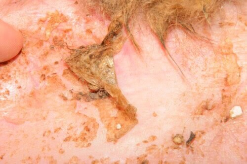
Feline pancreatic paraneoplastic alopecia
3.2 Thymoma-associated exfoliative dermatitis
Thymoma-associated exfoliative dermatitis presents a more encouraging picture. This paraneoplastic dermatosis shows as profuse non-pruritic scaling that starts on the head before spreading to the rest of the coat, and is often accompanied by generalised alopecia. Thoracic radiography reveals a space-occupying lesion in the cranial mediastinum, and skin histopathology shows a suggestive cell-poor interface dermatitis.
Unlike the previous syndromes, surgical thymectomy is curative here, and resection of the thymic mass leads, in most reported cases, to complete restoration of the skin architecture. The prognosis depends on whether the mass can be resected and on its histological benignity, with good prospects of cure when the thymoma is not invasive. This entity should be considered for any generalised non-pruritic scaling in an older cat, so different is its prognosis from that of the other feline paraneoplastic syndromes.
3.3 Feline “lung-digit” syndrome, a metastatic lesion not to be missed
One last syndrome deserves mention: “lung-digit” syndrome. This metastatic manifestation of a primary lung carcinoma sometimes first appears as a swollen digit, an ulcer or isolated lameness, before any respiratory signs. This misleading presentation, which first suggests a benign orthopaedic or traumatic problem, should raise the possibility of metastatic spread from the lung in an older cat and lead to thoracic imaging before any local treatment decision is made about the digital lesion.
This syndrome illustrates a cardinal principle of feline geriatric dermatology: any isolated digital lesion in an older cat, without an identified trauma, must lead to metastatic neoplasia being ruled out before it is treated as a benign local condition. Biopsy of the digital lesion together with thoracic imaging usually leads quickly to the diagnosis and avoids unnecessary, even harmful, local surgery in a patient whose overall prognosis is already compromised by pulmonary spread.
4 Towards dermatological vigilance in the older patient
At the end of this review of the main geriatric dermatoses of dogs and cats, one conclusion stands out: the skin of the older patient is not just a peripheral organ, but a sentinel organ, able to reveal early a systemic or neoplastic disease whose prognosis depends on prompt diagnosis. The ageing atopic dog therefore deserves closer dermatological follow-up, as a shift from long-stable pruritus to refractory signs should raise the possibility of underlying epitheliotropic cutaneous lymphoma. Likewise, footpad hyperkeratosis in an older dog, or non-pruritic ventral alopecia in an older cat, should lead to a thorough systemic work-up rather than simple local symptomatic treatment.
This vigilance is part of a comprehensive geriatric approach, in which every skin sign, however subtle, may reveal a systemic imbalance. Managing the older patient cannot rely on a fragmented view of the body: it requires constant integration of dermatological, biochemical, ultrasound and sometimes molecular data, in order to offer an overall diagnosis and care adapted to the patient’s comorbidities and comfort needs.
There are many avenues for research. Elucidating the molecular mechanisms linking chronic atopic inflammation to lymphomatous transformation is probably one of the most promising, as is the prospective evaluation of the long-term efficacy of rabacfosadine in the nodular-ulcerative tumour forms of epitheliotropic lymphoma, which are currently less well controlled than the diffuse erythrodermic forms. Molecular characterisation with the PARR test would benefit from greater standardisation, to reduce the false-negative rate linked to sample quality. Finally, a better understanding of the triggers and mechanisms of feline paraneoplastic syndromes, still poorly known because of their rarity, would probably improve their early recognition, a prerequisite for a better prognosis, particularly for paraneoplastic alopecia, whose outcome remains almost always fatal.
Reference
Oliveira A. Geriatric dermatology: uncovering skin disorders in senior patients. 35th European Veterinary Dermatology Congress. Practical Programme. 2025 Sep 11-13;Bilbao, Spain:24.
