

The paw pad is a specialised cutaneous structure designed for weight-bearing, capable of absorbing ground reaction forces by virtue of its distinctive micro-anatomy. This mechanical function accounts for both the considerable thickness of its cornified covering and the frequency of lameness when that covering is affected. Indeed, paw pad disorders rank among the common causes of lameness in the dog. What follows is an overview of the very numerous dermatoses affecting the paw pads in the dog.
The difficulty of the differential diagnosis of paw pad dermatoses lies in the fact that the elementary lesion of the paw pad, namely hyperkeratosis, has no orientating value in itself. Indeed, the same pedal hyperkeratosis is encountered in hereditary, metabolic, infectious, dysimmune and mechanical conditions, whose prognosis ranges from a purely cosmetic concern to a fatal outcome. Thus, the old term “hard pad disease”, which described paw pad thickening in canine distemper, was gradually abandoned, precisely because this presentation is also found in zinc-responsive dermatosis, hepatocutaneous syndrome and pemphigus foliaceus (Gröne et al., 2004a). In this review we shall discuss the principal paw pad dermatoses in the dog, with the aim of establishing the broadest possible differential diagnosis.
1 Anatomical and functional background
An understanding of paw pad dermatoses requires that we first recall what distinguishes this integument from haired skin. The paw pad consists of a thick stratified epithelium, endowed with an abundant cornified layer, deep epidermal ridges and tall dermal papillae that interdigitate extensively with the epidermis (Ninomiya et al., 2011). Its surface, smooth to the naked eye, is in reality formed of multiple conical papillae approximately 200 to 300 micrometres in height (Ninomiya et al., 2011). Histologically, the canine paw pad resembles human palmoplantar skin, with which it shares a thick cornified layer arranged in columns; it is distinguished, however, by particularly deep epidermal ridges (Bowden et al., 2009). This architecture illuminates one important point: the cornified layer of the paw pad, capable of absorbing forces reaching up to six times the body weight per limb, is also the most susceptible to fissuring as soon as its thickness and hardness increase at the expense of its pliability (Utzmann et al., 2025).
2 Diagnostic approach to paw pad disease
Since no pedal lesion is pathognomonic, reasoning takes precedence over visual recognition. A stepwise approach is therefore required: first characterising the elementary lesions, then placing them in their topographical and general context, before resorting to ancillary investigations. The history and full clinical examination are never incidental: foot dermatoses are most often a component of a more generalised dermatosis or a concurrent systemic condition, such that an examination limited to the foot alone risks overlooking the underlying disease (Jackson, 1999).
2.1 Elementary lesions and semiology
The lesions encountered on paw pads fall into a small number of types: fissures, erosions, ulcerations, crusts, margin hypertrophy, swelling and depigmentation. The vertical fissures that form on a hyperkeratotic pad are often painful and precede secondary inflammation and infection (Utzmann et al., 2025). Thus, the same hyperkeratosis may remain simple cornified thickening or may progress to deep cracks, and it is this evolution, more than the hyperkeratosis itself, that prompts the consultation. From a pathophysiological standpoint, thickening of the horn is accompanied by a loss of its plasticity: the cornified layer, thicker but less well hydrated, tolerates the shear stresses of weight-bearing poorly and fissures, opening the way to bacterial and yeast colonisation of the cracks (Utzmann et al., 2025).
2.2 Orientation by topography and context
The most useful discriminating factor is whether the disease is confined to the paw pads or is accompanied by lesions of the haired integument, mucocutaneous junctions or systemic signs. Indeed, metabolic, infectious and dysimmune dermatoses almost invariably produce lesions elsewhere than on the paw pads, or are accompanied by a poor general condition (Utzmann et al., 2025). However, two entities are exceptions: hereditary paw pad hyperkeratosis, which affects only the pads and begins in young animals, and idiopathic nasodigital hyperkeratosis, in which only the nasal planum is also involved (Utzmann et al., 2025). Strictly pedal involvement in a young dog of a predisposed breed therefore points immediately towards a primary keratinisation disorder, whereas involvement of several feet associated with systemic signs necessitates the investigation of a metabolic, infectious or immune cause. Whether the disease is mono- or multifocal constitutes a second axis of orientation: a lesion on a single pad suggests a corn, trauma or tumour, whereas symmetrical involvement of all four feet points towards an internal or hereditary cause (Hardy, 2017).
2.3 Role of biopsy and cytology
Histopathology occupies a central position. Parakeratotic hyperkeratosis, in which nuclei persist within the cornified layer, points towards zinc-responsive dermatosis (White et al., 2001) or hepatocutaneous syndrome (Gross et al., 1993), whilst orthokeratotic hyperkeratosis is more characteristic of hereditary forms (Drögemüller et al., 2014) and canine distemper (Koutinas et al., 2004). Cytology retains its value: the demonstration of acantholytic keratinocytes points towards pemphigus foliaceus, whilst the presence of amastigotes is conclusive for leishmaniosis. Two practical points deserve mention. Firstly, biopsy of the paw pad is technically demanding: the sample must come from a representative lesion, preferably non-ulcerated, be sufficiently deep to include the full thickness of the horn, and its healing, which is slow and painful in a weight-bearing area, requires a protective bandage and relative rest (Jackson, 1999). Technically, a 6 mm punch is suitable for most situations, but a wedge biopsy is preferable for deep, bullous or ulcerative lesions, as it allows both the margin of the lesion and the adjacent epidermis to be sampled whilst permitting careful suturing (Hnilica and Patterson, 2017). Secondly, surface cytology, performed by impression smear or swabbing of the cracks and interdigital spaces, clarifies the nature of secondary infection — cocci, bacilli or Malassezia — on which the choice of topical antimicrobial treatment depends (Ortalda et al., 2016). The work-up is completed, according to the clinical orientation, by bacteriological culture with sensitivity testing, fungal culture, a search for Demodex by skin scraping or trichogram, serology or PCR for leishmaniosis, and biochemical and hepatic ultrasonographic assessment when superficial necrolytic dermatitis is suspected (van den Broek and Horváth-Ungerböck, 2011; Burns DeMarle et al., 2021).
3 Primary keratinisation disorders
These conditions share a common feature: the lesion is confined to the paw pad, possibly associated with the nasal planum, without systemic signs. They arise from an intrinsic defect in cornification, often of genetic origin, and tend to manifest in young dogs.
3.1 Hereditary paw pad hyperkeratosis
Hereditary paw pad hyperkeratosis is transmitted as an autosomal recessive trait and was first reported in the Kromfohrländer and the Irish Terrier (Drögemüller et al., 2014). It results from a variant in the FAM83G gene, leading to the substitution of an arginine by a proline at position 52 of the PAWS1 protein (Drögemüller et al., 2014). This finding was confirmed by whole-genome sequencing of a family trio of Kromfohrländers, which identified the same variant as the sole candidate concordant with the phenotype (Sayyab et al., 2016). Mutations in FAM83G impair the interaction of the PAWS1 protein with casein kinase 1 alpha and attenuate the Wnt signalling pathway, a mechanism shared with palmoplantar keratodermas in humans (Wu et al., 2019).
Clinically, the condition begins in young dogs within the first six months of life and affects all four feet, with all pads becoming covered in a hard, dry and fissured horn that appears orthokeratotic on histological examination; the animals otherwise remain in good general health, but severe hyperkeratosis may be complicated by fissures, secondary infections and lameness (Drögemüller et al., 2014; Hardy, 2017). The monogenic aetiology permits a genetic test, enabling breeders to be advised against using carrier animals for breeding (Leeb et al., 2021). Related familial forms have moreover been described in the Dogue de Bordeaux and the Rottweiler (Utzmann et al., 2025; Leeb et al., 2021), and a variant in the GJB6 gene, encoding connexin 30, was recently reported in a Labrador Retriever with paw pad hyperkeratosis resembling human Clouston syndrome (Rietmann et al., 2026). These entities must be distinguished from the congenital “dry eye curly coat” syndrome of the Cavalier King Charles Spaniel, linked to a deletion of the FAM83H gene, in which paw pad hyperkeratosis is associated with keratoconjunctivitis sicca and nail dystrophy (Forman et al., 2012). The condition is not curable, and its management is purely symptomatic.
3.2 Idiopathic nasodigital hyperkeratosis
Idiopathic nasodigital hyperkeratosis corresponds to an alteration in cornification affecting the nasal planum and paw pads in dogs of various breeds; relatively common in older dogs, it is regarded as a senile modification of desquamation (Utzmann et al., 2025; Hardy, 2017). In its uncomplicated form it causes primarily a cosmetic concern; however, the prevention of fissures and secondary infections justifies management (Viaud et al., 2025). The hyperkeratosis is most noticeable at the margins of the pad, where the abrasive effect of weight-bearing is absent (Hardy, 2017). Treatment is based on the topical application of emollients and keratolytics. Indeed, a randomised placebo-controlled trial demonstrated, in idiopathic nasal hyperkeratosis, the efficacy of a balm containing essential oils and essential fatty acids applied daily (Catarino et al., 2018), and an open-label pilot study confirmed the good tolerability and clinical improvement obtained with an emollient balm (Viaud et al., 2025). These data relate to the nasal planum, and their extrapolation to the paw pad remains empirical.
3.3 Split paw pad dermatosis and cohesion abnormalities
Split paw pad dermatosis is characterised by horizontal fissuring and scaling of the pad horn, with frequent recurrence. The dominant signs are pain, lameness, pruritus and licking, with all four feet most commonly affected over the course of the disease; large-breed dogs weighing more than twenty kilograms are overrepresented (eight of thirteen dogs in the reference series, with a mean weight of 24.6 kilograms), the German Shepherd Dog is frequently mentioned, and seasonality as well as concurrent atopic dermatitis are noted in a proportion of cases (Utzmann et al., 2025). No single factor has been implicated, and several mechanisms must therefore be considered, including hypersensitivities, excessive shear forces, moisture, heat and overweight; no single treatment prevents relapses, and only barrier-supportive measures confer partial benefit, although a small subgroup does respond to anti-inflammatory treatment (Utzmann et al., 2025).
A genetically determined epidermal cohesion abnormality was identified in a family of German Shepherd Dogs presenting with intermittent pedal lesions and lameness. An in-frame 18-base-pair deletion in the KRT5 gene was demonstrated, associated with a cleavage plane within the stratum spinosum and stratum corneum, a finding likened to localised epidermolysis bullosa simplex in humans (Rietmann et al., 2024). This observation illustrates the fact that a structural defect in a keratin filament can manifest as fragility of the paw pad horn.
4 Metabolic and nutritional dermatoses
Unlike primary keratinisation disorders, these dermatoses are the consequence of a systemic, hepatic, pancreatic or nutritional disturbance. Pedal involvement is therefore rarely isolated, and its recognition is valuable in that it opens the way to investigation of the underlying condition.
4.1 Superficial necrolytic dermatitis (formerly hepatocutaneous syndrome)
Superficial necrolytic dermatitis, also termed hepatocutaneous syndrome or metabolic epidermal necrosis, is a rare condition of grave prognosis affecting middle-aged to elderly dogs, with no established breed or sex predisposition (Cellio and Dennis, 2005). It presents as an erosive, crusting and squamous dermatosis with a symmetrical distribution involving the face, distal limbs, inguinal region, mucocutaneous junctions and pressure points; paw pad involvement, consisting of hyperkeratosis, fissuring and ulceration, is almost invariably present and was recorded in twenty-one of twenty-two dogs in a seminal series (Gross et al., 1993). The history commonly includes lethargy, anorexia, weight loss and a painful gait (Cellio and Dennis, 2005).
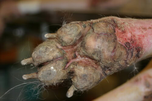
Severe form of superficial necrolytic dermatitis
Pathophysiologically, the condition is most often associated with severe vacuolar hepatopathy, and less frequently with a pancreatic neuroendocrine tumour or glucagonoma (Gross et al., 1990; Papadogiannakis et al., 2009). Prolonged administration of phenobarbitone, or phenytoin, has also been cited among triggering contexts (Papadogiannakis et al., 2009; van den Broek and Horváth-Ungerböck, 2011), as has copper-associated hepatitis (Talbot et al., 2022). Cutaneous histology is highly suggestive: parakeratosis, intercellular and intracellular oedema of the upper half of the epidermis and hyperplasia of the basal layers together form the classic layered appearance (Gross et al., 1993). Given the non-specific nature of the clinical signs, diagnosis rests on targeted investigations: marked plasma hypoaminoacidaemia was present in all cases in a large review, and abdominal ultrasonography showed a honeycomb appearance of the liver in almost all affected dogs, permitting a minimally invasive diagnostic approach based on the amino acid profile complemented by imaging (Burns DeMarle et al., 2021).
Treatment remains disappointing and essentially palliative: amino acid supplementation by intravenous or oral routes, high-quality protein intake, and, when copper-associated hepatitis is confirmed, copper chelation (Talbot et al., 2022). Zinc and essential fatty acid supplementation, together with topical antiseptic and keratolytic care of pedal lesions, add to the comfort of the animal (van den Broek and Horváth-Ungerböck, 2011). Nevertheless, the prognosis remains grave, with most dogs dying or being euthanised within months of the onset of signs (Cellio and Dennis, 2005).
4.2 Zinc-responsive dermatoses
Zinc-responsive dermatoses fall into two forms. The first is a familial form in northern breeds, foremost among which are the Siberian Husky and the Alaskan Malamute, in which a defect in zinc absorption or metabolism is responsible; the second affects growing puppies fed a zinc-deficient or unbalanced diet (Colombini, 1999). Periocular crusting is the most frequent sign, and parakeratosis is found on biopsy in all dogs in a series of forty-one cases dominated by the Siberian Husky (White et al., 2001). Involvement of the paw pads and pressure points is possible, with lesions often beginning unilaterally before becoming symmetrical, with a seasonal component (Colombini et al., 1997).
Treatment is based on oral zinc supplementation. An initial dose of 2 to 3 mg/kg per day of elemental zinc is recommended in the series of White et al. (2001), whilst Colombini et al. (1997) suggest starting at 1 mg/kg per day and increasing gradually in the absence of a response. The familial form requires lifelong supplementation, and relapses occur readily when a dose is missed or the dosage is reduced (Colombini et al., 1997). Furthermore, localised parakeratotic hyperkeratoses resembling zinc-responsive dermatosis have recently been described on the pinnae of the Boston Terrier and the French Bulldog, with partial response to supplementation, without any difference in tissue zinc concentration compared with controls (Lee et al., 2016; Dubin et al., 2025).
4.3 Lethal acrodermatitis of the Bull Terrier
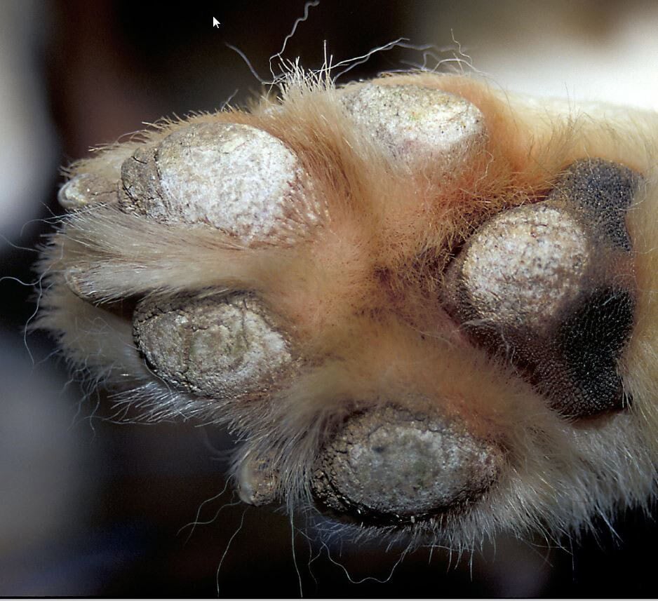
Lethal acrodermatitis is an autosomal recessive genodermatosis of the Bull Terrier and Miniature Bull Terrier, linked to a variant in the MKLN1 gene (Bauer et al., 2018). The clinical picture combines growth retardation, splayed digits, dermatosis of the face and feet and immune deficiency; in older animals paronychia, nail involvement and paw pad hyperkeratosis appear, the latter being more severe once the puppy exceeds six months of age (McEwan et al., 2000; Jezyk et al., 1986). Histology reveals a parakeratotic hyperkeratosis resembling zinc deficiency, but, unlike zinc-responsive dermatoses, the condition does not respond to zinc supplementation and carries a median survival of approximately seven months (Jezyk et al., 1986). Genetic testing now enables the identification of carriers and the avoidance of affected offspring (Bauer et al., 2018).
4.4 Other nutritional deficiencies and imbalances
Beyond zinc-responsive dermatoses, pedal cornification may suffer from broader nutritional imbalances, notably in poorly formulated home-prepared diets or atypical feeding regimens. The older concept of “generic dog food dermatosis”, reported in puppies fed low-grade food, represents the archetype and is linked, in part, to a deficiency in zinc intake or bioavailability (Utzmann et al., 2025). Essential fatty acid deficiency also impairs barrier function and horn quality; their supplementation is proposed in the long-term management of several pedal dermatoses, including symmetrical lupoid onychodystrophy (van den Broek and Horváth-Ungerböck, 2011). These considerations justify including a dietary history in the work-up of pedal hyperkeratosis in a young dog, before concluding that a hereditary form is present.
5 Infectious and parasitic dermatoses
The infectious aspect of pedal disease falls into two categories. On the one hand, two conditions stand out for the consistency with which they affect the paw pad itself: canine distemper, whose pad thickening long gave it its common name, and leishmaniosis, in which nasodigital hyperkeratosis accompanies onychogryphosis; in both cases, pedal involvement is part of a systemic picture that prompts their suspicion.
5.1 Canine distemper
Hyperkeratosis of the paw pads and nasal planum, referred to as “hard pad disease”, constitutes a late and infrequent sequela of infection with canine distemper virus (Koutinas et al., 2004; Gröne et al., 2004a). Histologically, it produces an orthokeratotic hyperkeratosis associated with acanthosis and thickening of the epidermal ridges; viral antigen localises preferentially to the stratum spinosum and stratum granulosum as well as to the cells of the eccrine sweat glands (Koutinas et al., 2004; Gröne et al., 2004a), and is accompanied by a disorder of keratinocyte differentiation, evidenced by modification of cytokeratin expression (Gröne et al., 2004b). Inclusion bodies and ballooning degeneration, formerly regarded as characteristic, are in fact inconstant in naturally infected paw pads (Koutinas et al., 2004). Beyond the nasodigital regions, hyperkeratosis may affect other sites, such as the nasal planum, the periocular area or the ventral abdomen (Areco et al., 2022).
Pedal involvement fits into the general picture of canine distemper, with its respiratory, digestive and neurological components, which facilitates its attribution. Demonstration of the virus in the paw pad epidermis has moreover been proposed as a means of antemortem diagnosis (Gröne et al., 2004a).
5.2 Leishmaniosis
In canine leishmaniosis due to Leishmania infantum, cutaneous lesions constitute the most frequent clinical manifestation, and exfoliative dermatitis represents its dominant form (Saridomichelakis et al., 2014). Nasodigital hyperkeratosis and onychogryphosis are among the characteristic signs (Koutinas et al., 2014), alongside ulceration of the extremities and squamous scaling (Ferrer et al., 1988). Definitive diagnosis rests on the combination of the macroscopic appearance, exclusion of the main differential diagnoses, histopathology, demonstration of the parasite and complete response to antileishmanial treatment (Saridomichelakis et al., 2014). However, species such as Leishmania major may produce ulcerative lesions of the muzzle, feet and paw pads, sometimes without seropositivity, which then necessitates molecular biological techniques (Baneth et al., 2016). Furthermore, ischaemic dermatopathy and vasculitis lesions have been reported in association with leishmaniosis, of which they may constitute a manifestation (García et al., 2025). Given its Mediterranean distribution, this hypothesis must be considered in the light of the animal’s geographical origin and any stays in an enzootic area.
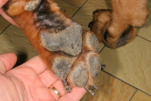
Paw pad thickening in a dog with leishmaniosis
6 Dysimmune dermatoses
Immune-mediated dermatoses occupy a particular position, since paw pad involvement is both frequent and sometimes the initial presentation. Pemphigus foliaceus is the most common representative, whilst cutaneous lupus, drug reactions and vasculopathies complete the autoimmune and ischaemic spectrum of pedal disease.
6.1 Pemphigus foliaceus
Pemphigus foliaceus is the most common autoimmune dermatosis in the dog. It produces pustules, crusts, erosions, scales and alopecia affecting principally the muzzle, face, inner surface of the pinnae and trunk (Ihrke et al., 1985b; Mueller et al., 2006). Paw pad involvement is common, present in nearly one third of cases, and manifests as erythematous swelling of the margins, fissuring and villous hypertrophy (Ihrke et al., 1985b; van den Broek and Horváth-Ungerböck, 2011). A pemphigus foliaceus limited to the paw pads has indeed been described, producing a picture of hyperkeratinisation and villous hypertrophy that misleadingly resembles “hard pad disease” (Ihrke et al., 1985a). The coexistence of vasculitis lesions prolongs the time to remission (Zhou et al., 2021).
Diagnosis relies on cytology, which demonstrates acantholytic keratinocytes, and on histopathology, which shows subcorneal or intragranular pustules rich in acanthocytes. The major autoantigen is desmocollin 1 (Bizikova et al., 2011; Bizikova et al., 2022). Treatment is based on immunosuppressive-dose corticosteroid therapy, possibly combined with azathioprine, the combination having failed to demonstrate superiority over corticosteroids alone in a series of ninety-one dogs (Mueller et al., 2006). Oclacitinib has moreover been proposed as an emerging option (Jordan et al., 2024).
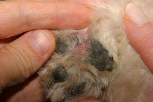
Pedal lesions in a dog with pemphigus foliaceus
6.2 Cutaneous lupus erythematosus
The spectrum of canine cutaneous lupus erythematosus has broadened considerably since the initial description of discoid lupus, and a canine adaptation of the Gilliam and Sontheimer classification now distinguishes several variants: vesicular cutaneous lupus, exfoliative cutaneous lupus, mucocutaneous lupus and facial or generalised discoid lupus (Olivry et al., 2018). Several of these forms affect the extremities: vesicular cutaneous lupus produces annular and polycyclic erosions on glabrous areas, including the abdomen and inner thighs, whilst mucocutaneous lupus affects the junctions and may extend to the extremities (Olivry et al., 2018). Paw pad involvement is possible in the context of systemic lupus, whose cutaneous manifestations, which are rarer, appear at the margins of the spectrum (Olivry et al., 2018). Most of these variants carry a favourable prognosis once diagnosed, which justifies recognising them in order to institute appropriate immunomodulatory treatment at an early stage.
6.3 Drug reactions and erythema multiforme
Drug-induced cutaneous reactions and erythema multiforme deserve a place in the differential diagnosis of acute, erosive or ulcerative pedal disease. Canine erythema multiforme covers a spectrum ranging from erosive and vesiculobullous forms to a recently individualised hyperkeratotic variant: described in seventeen dogs, most often middle-aged to older males, this variant produces erythematous annular and linear macules and plaques covered with firm, adherent crusts, with panepidermal cytotoxic dermatitis and ortho- and parakeratotic hyperkeratosis on histology; it is distinguished from classic erosive erythema multiforme and does not respond to antimicrobials, but does respond to immunosuppressants in just over half of cases (Banovic et al., 2023). The severe forms include Stevens-Johnson syndrome and toxic epidermal necrolysis, acute and grave dermatoses in which pedal involvement, characterised by ulceration and pain, manifests as widespread epidermal detachment often triggered by a drug. Recognition of these entities requires discontinuation of the suspected drug and supportive care, and clearly distinguishes their prognosis from that of chronic hyperkeratoses.
Alongside strictly immune drug reactions, certain pedal lesions arise from direct iatrogenic injury. Systemic retinoids, such as acitretin, count among their adverse effects cracking and fissuring of the paw pads, which form part of the picture of cheilitis and exfoliative dermatitis (Koch et al., 2012). Tyrosine kinase inhibitors used in oncology, including toceranib, expose the patient to lameness, skeletal pain, dermatitis, pruritus and pigmentation disorders (Koch et al., 2012).
6.4 Vasculopathies and ischaemic dermatopathies
Familial cutaneous vasculopathy of the German Shepherd Dog illustrates ischaemic involvement of the paw pad. Described in puppies, it combines swelling, depigmentation and ulceration of the paw pads with crusting of the ear tips and tail, focal depigmentation of the nasal planum, and fever and lethargy (Weir et al., 1994). Histology reveals a multifocal nodular dermatitis in which neutrophils and mononuclear cells surround foci of dermal collagenolysis, accompanied by degenerative and inflammatory vascular lesions; the mode of transmission is autosomal recessive (Weir et al., 1994). A case associating this vasculopathy with generalised demodicosis has moreover been reported (Fondati et al., 1998). Ulceration of the central pads constitutes a characteristic presenting sign. More generally, ischaemic dermatopathy and vasculitis may complicate other conditions, foremost among which is leishmaniosis (García et al., 2025), and should be considered in the presence of pedal ulceration with well-defined edges accompanied by distal extremity lesions.
7 Mechanical, traumatic and neoplastic conditions
This final group brings together conditions whose common feature is that they do not result from a systemic disorder, but from physical injury, repeated mechanical stress or neoplastic proliferation. Their recognition is important, as it entirely redirects management, from surgical intervention to simple pad protection.
7.1 Corns and paw pad keratomas
A corn presents as a focal, circular, firm and hyperkeratotic lesion, typically situated at the centre of the digital pads (Utzmann et al., 2025). It is found preferentially on the third and fourth digits and the thoracic limbs, affects mainly Greyhounds and related breeds, and predominantly males (Guilliard et al., 2010). Its frequency is by no means negligible in this population: reported at between 2.4 and 5.9% in retired racing Greyhounds, it constitutes the most common dermatological condition in these dogs (Guilliard and Doughty, 2022). It causes chronic lameness that may be severe, aggravated on hard ground, and mediolateral digital pressure consistently elicits pain (Guilliard and Doughty, 2022). The proposed mechanisms are mechanical, whether traumatic or due to repeated pressure, or the penetration of a foreign body; the viral hypothesis, for a time supported by the demonstration of a papillomavirus in two Greyhounds (Anis et al., 2016), is now considered unlikely, as infection was not found in a series of eighteen Greyhounds nor on histological examination of more than one thousand corns (Guilliard and Doughty, 2022). Treatment remains difficult: surgical excision carries a recurrence rate exceeding half of cases, distal digital ostectomy is considered only in carefully selected cases, and tenotomy of the superficial digital flexor tendon, by modifying the loading of the phalanx, provides immediate pain relief (Guilliard et al., 2010; Guilliard and Doughty, 2022; Martinez et al., 2021).
7.2 Burns and physicochemical injuries
Injury to a single isolated pad may also result from trauma by an object, such as a piece of glass, and as a rule heals without complication in an otherwise healthy animal within a few weeks, depending on the extent of the wound (Utzmann et al., 2025). In contrast, simultaneous involvement of several pads suggests an environmental insult: thermal burns from a hot surface, such as a road or beach in summer, chemical burns from irritants, or barrier disruption caused by road salt and winter moisture (Utzmann et al., 2025). The topography and whether the disease is mono- or multifocal therefore remain, here too, key orientating features. In working and sporting dogs, intense use of the paw pads is a specific risk factor: in sled dogs, exercise-related pododermatitis is common, and an emollient balm applied before and after exercise significantly reduces the occurrence of erythema, abrasions and cracks (Bouvier et al., 2020).
7.3 Paw pad tumours
Tumours of the paw pad proper are rare but must be included in the differential diagnosis of a solitary pedal nodule or thickening. Malignant melanoma of the paw pad, a poorly documented entity, behaves aggressively: in a multicentre series of twenty cases, the overall metastatic rate exceeded half of the dogs and the median survival was approximately two hundred and forty days (Jeon et al., 2022). A primary cutaneous ganglioneuroblastoma of the paw pad has moreover been described, manifesting as lameness and pedal licking associated with thickening of a digital pad (Salvadori et al., 2019). Thus, any nodular, unilateral and persistent pedal lesion warrants histological investigation before being attributed to a benign cause.
7.4 Calcinosis and mineral deposits
Cutaneous calcinosis deserves consideration when a firm nodule is found at pressure points or in the pedal region. Circumscribed calcinosis is a nodule resulting from dystrophic calcification, with lesions arising principally at sites of repeated or previous trauma: pressure points, paw pads and areas of injury (Hnilica and Patterson, 2017). It tends to affect young, rapidly growing dogs, with the German Shepherd Dog, the Boston Terrier and the Boxer being predisposed; it produces firm, well-circumscribed nodules, generally solitary, measuring 0.5 to 7 cm, situated in the subcutaneous tissue, from which a whitish, chalk-like or pasty material may exude (Hnilica and Patterson, 2017). These nodules are non-painful, except precisely when they are located within the paw pads. Diagnosis combines cytology, which shows calcium fragments and granulomatous inflammation, histopathology, and, when necessary, radiography to demonstrate calcific deposits lying deep within the metacarpal or metatarsal pads (Hnilica and Patterson, 2017). Surgical excision is curative. It should be distinguished from diffuse cutaneous calcinosis, which is observed principally in the context of hyperadrenocorticism (Miller et al., 2013).
8 Symptomatic management common to hyperkeratosis and fissures
Whatever the underlying cause, fissured pedal hyperkeratosis calls for a transversal symptomatic approach aimed at softening the horn, restoring the barrier and protecting the pad from weight-bearing, whilst aetiological treatment, where available, is conducted in parallel. This practical dimension, often the only one available in incurable hereditary or idiopathic forms, merits consideration in its own right.
The first lever is keratolytic and moisturising. Urea at high concentration, from forty to fifty per cent, is a keratolytic of choice for hyperkeratotic dermatoses: it dissolves the proteins of the intercellular matrix, detaches the cornified layer and simultaneously moisturises the underlying epidermis, whilst also increasing the penetration of any associated active ingredients (Starace et al., 2020). Salicylic acid, at concentrations of approximately ten per cent, combined with regular trimming of excess horn, markedly reduces paw pad hyperkeratosis and the accompanying lameness (De Lucia et al., 2019). Emollient balms based on essential fatty acids and essential oils, applied daily, improve the condition of the horn and prevent fissuring (Catarino et al., 2018; Viaud et al., 2025).
The second lever is cutaneous barrier support and mechanical protection. In split paw pad dermatosis as in hereditary hyperkeratoses, protective measures for the pad — boots and bandages — combined with barrier-reinforcing treatment, confer benefit where no single treatment prevents relapses (Utzmann et al., 2025). In working dogs, the preventive application of a balm before exercise limits the development of lesions (Bouvier et al., 2020).
The third lever addresses secondary infection and inflammation of the fissures. Surface cytology guides the choice of topical antiseptic or antifungal, whose efficacy against bacterial and yeast burdens on the foot has been demonstrated (Ortalda et al., 2016); antiseptic and keratolytic shampoos and soaking of crusted lesions are useful complements to this care (van den Broek and Horváth-Ungerböck, 2011). Given the chronic and relapsing nature of most of these conditions, owner education, explanation of the importance of daily topical care and a scheduled reassessment programme are essential to compliance and long-term outcome.
Pain and lameness management must not be neglected, even though it is the predominant reason for the consultation. Deep fissures and burns are painful and warrant specific analgesia and relative rest; in the particular case of the corn, when conservative and surgical measures fail, tenotomy of the superficial digital flexor tendon provides immediate relief by modifying phalangeal loading (Martinez et al., 2021; Guilliard and Doughty, 2022).
Conclusion
Paw pad dermatoses share a common final pathway — hyperkeratosis — which renders them clinically similar and diagnostically deceptive. Thus, reasoning takes precedence over recognition: characterising the elementary lesions, assessing whether the disease extends beyond the pad, distinguishing involvement of the cornified pad from that of the interdigital spaces, placing the picture in its context of breed, age, geographical origin and systemic signs, then confirming by histopathology and, as appropriate, genetics, serology, molecular biology or hepatic work-up. Ultimately, the prognosis ranges from a purely cosmetic concern, as in idiopathic nasodigital hyperkeratosis, to the fatal outcome of hepatocutaneous syndrome, which means that diagnostic rigour has direct prognostic and therapeutic implications. When aetiological treatment is unavailable, rigorous symptomatic management of the horn, the barrier and pain remains, in itself, a tangible benefit for the animal.
References
Anis EA, Frank LA, Francisco R, Kania SA. Identification of canine papillomavirus by PCR in Greyhound dogs. PeerJ. 2016.
Areco WC, et al. Macroscopic distribution, histopathology and viral antigen expression in dogs with canine distemper virus-induced hyperkeratosis in nasodigital and other regions. J Comp Pathol. 2022.
Baneth G, et al. Leishmania major infection in a dog with cutaneous manifestations. Parasit Vectors. 2016.
Banovic F, Olivry T, Artlet B, et al. Hyperkeratotic erythema multiforme variant in 17 dogs. Vet Dermatol. 2023;34(2):125-133. https://doi.org/10.1111/vde.13141
Bauer A, et al. MKLN1 splicing defect in dogs with lethal acrodermatitis. PLoS Genet. 2018;14:e1007264. https://doi.org/10.1371/journal.pgen.1007264
Bizikova P, Linder KE, Olivry T. Immunomapping of desmosomal and nondesmosomal adhesion molecules in healthy canine footpad, haired skin and buccal mucosal epithelia: comparison with canine pemphigus foliaceus serum immunoglobulin G staining patterns. Vet Dermatol. 2011;22(2):132-142. https://doi.org/10.1111/j.1365-3164.2010.00924.x
Bizikova P, et al. Trunk-dominant and classic facial pemphigus foliaceus in dogs: comparison of anti-desmocollin-1 and anti-desmoglein-1 autoantibodies and clinical presentations. Vet Dermatol. 2022.
Bouvier A, Thorin C, Pouliquen H, Bourdeau P. Evaluation of a topical balm on foot pad lesions of sled dogs during intensive training [abstract]. Vet Dermatol. 2020;31(S1):6-102. https://doi.org/10.1111/vde.12907
Bowden PE, Henderson H, Reilly JD. Defining the complex epithelia that comprise the canine claw with molecular markers of differentiation. Vet Dermatol. 2009;20(5-6):347-359. https://doi.org/10.1111/j.1365-3164.2009.00851.x
Burns DeMarle K, Webster CRL, Penninck D, Ferrer L. Approach to the diagnosis of hepatocutaneous syndrome in dogs: a retrospective study and literature review. J Am Anim Hosp Assoc. 2021;57(1):15-25. https://doi.org/10.5326/JAAHA-MS-7072
Catarino M, et al. Control of canine idiopathic nasal hyperkeratosis with a natural skin restorative balm: a randomized double-blind placebo-controlled study. Vet Dermatol. 2018.
Cellio LM, Dennis J. Canine superficial necrolytic dermatitis. Compend Contin Educ Pract Vet. 2005;27:820-825.
Colombini S, et al. Zinc-responsive dermatosis in northern-breed dogs: 17 cases (1990-1996). J Am Vet Med Assoc. 1997.
Colombini S. Canine zinc-responsive dermatosis. Vet Clin North Am Small Anim Pract. 1999.
De Lucia M, Angileri M, Bauer A, et al. X-linked cutaneous mosaicism in a dog. Vet Dermatol. 2019;30(4):361-362. https://doi.org/10.1111/vde.12748
Drögemüller M, et al. A mutation in the FAM83G gene in dogs with hereditary footpad hyperkeratosis (HFH). PLoS Genet. 2014;10:e1004370. https://doi.org/10.1371/journal.pgen.1004370
Dubin RJ, et al. Pinnal parakeratotic hyperkeratosis consistent with zinc-responsive dermatosis in 16 French bulldogs. Vet Dermatol. 2025.
Fenet M, Layssol-Lamour C, Pressanti C, et al. Ultrasonographic findings may be useful for differentiating interdigital abscesses secondary to migrating grass awns and interdigital furunculosis in dogs. Vet Radiol Ultrasound. 2023;64(5):920-929. https://doi.org/10.1111/vru.13273
Ferrer L, et al. Skin lesions in canine leishmaniasis. J Small Anim Pract. 1988.
Fondati A, et al. Familial cutaneous vasculopathy and demodicosis in a German shepherd dog. J Small Anim Pract. 1998.
Forman OP, et al. Parallel mapping and simultaneous sequencing reveals deletions in BCAN and FAM83H associated with discrete inherited disorders in a domestic dog breed. PLoS Genet. 2012;8:e1002462. https://doi.org/10.1371/journal.pgen.1002462
García N, et al. Clinical, immunological and pathological characteristics of ischemic dermatopathy in dogs with leishmaniosis. Pathogens. 2025.
Gröne A, Doherr MG, Zurbriggen A. Canine distemper virus infection of canine footpad epidermis. Vet Dermatol. 2004a;15(3):159-167. https://doi.org/10.1111/j.1365-3164.2004.00384.x
Gröne A, Doherr MG, Zurbriggen A. Up-regulation of cytokeratin expression in canine distemper virus-infected canine footpad epidermis. Vet Dermatol. 2004b;15(3):168-174. https://doi.org/10.1111/j.1365-3164.2004.00385.x
Gross TL, et al. Glucagon-producing pancreatic endocrine tumors in two dogs with superficial necrolytic dermatitis. J Am Vet Med Assoc. 1990.
Gross TL, et al. Superficial necrolytic dermatitis (necrolytic migratory erythema) in dogs. Vet Pathol. 1993.
Guilliard MJ, et al. Corns in dogs; signalment, possible aetiology and response to surgical treatment. J Small Anim Pract. 2010;51(3):162-168. https://doi.org/10.1111/j.1748-5827.2010.00892.x
Guilliard MJ, Doughty RW. Superficial digital flexor tendonectomy for the treatment of corns in sighthounds. Vet Dermatol. 2022;33(6):581-586. https://doi.org/10.1111/vde.13117
Hardy J. Skin diseases affecting the nasal planum and footpads of dogs. In Pract. 2017;39(5):203-213. https://doi.org/10.1136/inp.j1762
Hnilica KA, Patterson AP. Skin diseases of the dog and cat. 3rd ed. St Louis: Elsevier Saunders; 2017.
Ihrke PJ, et al. Pemphigus foliaceus of the footpads in three dogs. J Am Vet Med Assoc. 1985a;186(1):67-69.
Ihrke PJ, Stannard AA, Ardans AA, Griffin CE. Pemphigus foliaceus in dogs: a review of 37 cases. J Am Vet Med Assoc. 1985b;186(1):59-66.
Jackson H. Common cutaneous diseases of the canine foot. In Pract. 1999;21(2):54-61. https://doi.org/10.1136/inpract.21.2.54
Jeon MD, et al. Multi-institutional retrospective study of canine foot pad malignant melanomas: 20 cases. Vet Comp Oncol. 2022. https://doi.org/10.1111/vco.12846
Jezyk PF, et al. Lethal acrodermatitis in bull terriers. J Am Vet Med Assoc. 1986.
Jordan TJM, et al. Canine and feline pemphigus foliaceus: an update on pathogenesis and treatment. Vet Clin North Am Small Anim Pract. 2024.
Koch SN, Torres SMF, Plumb DC. Canine and feline dermatology drug handbook. Chichester: Wiley-Blackwell; 2012.
Koutinas AF, et al. Histopathology and immunohistochemistry of canine distemper virus-induced footpad hyperkeratosis (hard pad disease) in dogs with natural canine distemper. Vet Pathol. 2004.
Koutinas AF, et al. Pathologic mechanisms underlying the clinical findings in canine leishmaniosis due to Leishmania infantum/chagasi. Vet Pathol. 2014.
Lange A, Mayer U, Bensignor E, et al. A blinded randomised split-body clinical trial evaluating the effect of fluorescent light energy on antimicrobial management of canine interdigital furunculosis. Vet Dermatol. 2025;36(3):291-301. https://doi.org/10.1111/vde.13340
Lee FF, et al. Localized parakeratotic hyperkeratosis in sixteen Boston terrier dogs. Vet Dermatol. 2016.
Leeb T, et al. Genetics of inherited skin disorders in dogs. Vet J. 2021.
Marchegiani A, Fruganti A, Gavazza A, Spaterna A, Cerquetella M. Fluorescence biomodulation for canine interdigital furunculosis: updates for once-weekly schedule. Front Vet Sci. 2022;9:880349. https://doi.org/10.3389/fvets.2022.880349
Martinez N, McDonald B. Superficial digital flexor tenotomy immediately relieves pain associated with paw pad keratomas in three dogs: a case report. Jpn J Vet Dermatol. 2021;27(3).
McEwan NA, et al. Diagnostic features, confirmation and disease progression in 28 cases of lethal acrodermatitis of bull terriers. J Small Anim Pract. 2000.
Miller WH, Griffin CE, Campbell KL. Muller and Kirk’s small animal dermatology. 7th ed. St Louis: Elsevier Mosby; 2013.
Mueller RS, et al. Pemphigus foliaceus in 91 dogs. J Am Anim Hosp Assoc. 2006.
Ninomiya H, et al. Functional anatomy of the footpad vasculature of dogs: scanning electron microscopy of vascular corrosion casts. Vet Dermatol. 2011;22(6):475-481. https://doi.org/10.1111/j.1365-3164.2011.00976.x
Olivry T, Linder KE, Banovic F. Cutaneous lupus erythematosus in dogs: a comprehensive review. BMC Vet Res. 2018;14(1):132. https://doi.org/10.1186/s12917-018-1446-8
Ortalda C, Noli C, Cena T. Efficacy of an ethanol/guar/triclosan/glycerine gel on bacteria and yeast loads in canine pododermatitis: a pilot study. J Small Anim Pract. 2016;57(4):205-209. https://doi.org/10.1111/jsap.12446
Papadogiannakis E, et al. Superficial necrolytic dermatitis in a dog associated with hyperplasia of pancreatic neuroendocrine cells. J Small Anim Pract. 2009.
Rietmann SJ, et al. KRT5 in-frame deletion in a family of German shepherd dogs with split paw pad disease resembling localized epidermolysis bullosa simplex in human patients. Anim Genet. 2024.
Rietmann SJ, et al. GJB6 missense variant in a Labrador retriever with paw pad hyperkeratosis. Anim Genet. 2026.
Salvadori C, et al. Footpad peripheral ganglioneuroblastoma in a dog. Vet Dermatol. 2019.
Saridomichelakis MN, Koutinas AF. Cutaneous involvement in canine leishmaniosis due to Leishmania infantum (syn. L. chagasi). Vet Dermatol. 2014;25(2):61-e22. https://doi.org/10.1111/vde.12105
Sayyab S, et al. Whole-genome sequencing of a canine family trio reveals a FAM83G variant associated with hereditary footpad hyperkeratosis. G3 (Bethesda). 2016;6:521-527. https://doi.org/10.1534/g3.115.025643
Starace M, Alessandrini A, Piraccini BM. Clinical evidences of urea at high concentration on skin and annexes. Int J Clin Pract. 2020;74(S187):e13740. https://doi.org/10.1111/ijcp.13740
Talbot C, et al. Treatment of superficial necrolytic dermatitis with copper chelation in a dog with copper-associated hepatitis. J Am Anim Hosp Assoc. 2022.
Utzmann M, Bettenay S, Mueller RS, Mayer U. Clinical signs and treatment responses of 14 dogs with split paw pad dermatosis. Vet Dermatol. 2025;36(6):903-913. https://doi.org/10.1111/vde.70007
van den Broek A, Horváth-Ungerböck C. Pedal dermatitis Part 2: Canine pododermatitis. Companion Anim. 2011;16(2):41-46. https://doi.org/10.1111/j.2044-3862.2010.00022.x
Viaud S, et al. Evaluation of the interest in and tolerance of a topical emollient in the management of canine nasal hyperkeratosis: an open-label, prospective, uncontrolled pilot study. Vet Sci. 2025.
Waisglass S. Claw disease in the dog: does your patient have symmetrical lupoid onychodystrophy (SLO)? Can Vet J. 2018;59(7):796-798.
Weir J, et al. Familial cutaneous vasculopathy of German shepherds: clinical, genetic and preliminary pathological and immunological studies. Can Vet J. 1994.
White SD, et al. Zinc-responsive dermatosis in dogs: 41 cases and literature review. Vet Dermatol. 2001.
Wu KZL, et al. Pathogenic FAM83G palmoplantar keratoderma mutations inhibit the PAWS1:CK1α association and attenuate Wnt signalling. Wellcome Open Res. 2019.
Zhou Z, et al. Clinical presentation, treatment and outcome in dogs with pemphigus foliaceus with and without vasculopathic lesions: an evaluation of 41 cases. Vet Dermatol. 2021.
