On the occasion of GEDAC’s last Annual Days, in Ajaccio, in June 2024, our colleague, Vincent Bruet, DipECVD, provided a comprehensive update on the use of antifungals in the treatment of canine and feline dermatophytosis.
Dermatophytosis represents the most frequently encountered fungal skin infections in companion animal veterinary medicine. These conditions, caused by keratinophilic fungi, pose particular therapeutic challenges due to their contagious nature and zoonotic potential. In the current context, where the medicalization of companion animals is constantly progressing, an update of knowledge on therapeutic strategies is essential.
Epidemiology and zoonotic aspects
In routine clinical practice, dermatophytes are subject to a constantly evolving classification. Although the genera Microsporum and Trichophyton remain the most commonly used, a new nomenclature now integrates the genus Nannizzia. The four predominant species in infections of domestic carnivores are Microsporum canis, a major zoophilic agent, particularly in cats where the prevalence reaches 6% of the feline population in consultation, Trichophyton mentagrophytes, also zoophilic, Nannizzia gypsea (formerly Microsporum gypseum), a geophilic agent, and Nannizzia persicolor (formerly Microsporum persicolor).
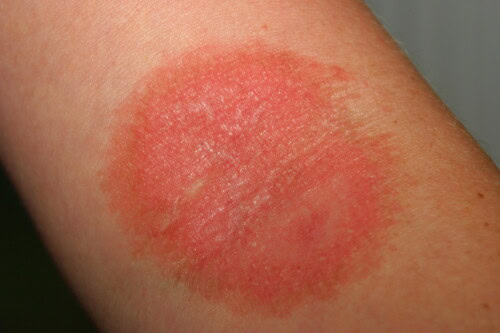
The zoonotic importance of these infections should not be underestimated. Studies reveal significant human contamination rates: 30% of infected cat owners and 15% of infected dog owners develop lesions. Transmission can be direct (contact with an infected animal) or indirect via the environment, with spore survival in the soil potentially reaching several months.
Photo 1: We must not forget that ringworm can be transmitted to humans.
Seasonality plays an important role in the epidemiology of certain species. While M. canis does not show a marked seasonal variation, T. mentagrophytes exhibits an autumnal recrudescence, correlated with the multiplication of small rodents which constitute its natural reservoir.
Hair invasion is mainly ectothrix in carnivores, with filaments inside the hair and arthrospores on the surface. The spore, showing a particular affinity for keratin, penetrates the hair follicle down to the isthmic zone, where it finds optimal conditions for its growth. This process leads to a weakening of the hair which, by breaking, releases new spores, thus perpetuating the infectious cycle according to a characteristic centripetal evolution. The incubation period varies from 10 to 30 days.
Diagnostic tools and therapeutic monitoring
Multimodal diagnostic approach
The diagnosis of dermatophytosis relies on a combination of complementary examinations. Wood’s lamp, while useful for screening M. canis, only detects about 50% of positive cases due to the variable production of fluorescent metabolites. Trichogram offers a rapid approach but its sensitivity strongly depends on the examiner’s experience.
Mycological culture remains the reference method, with results available in 1 to 3 weeks. Samples can be taken by skin scraping, targeted epilation under Wood’s lamp, or brushing the coat for asymptomatic infections or therapeutic monitoring.
The major innovation lies in the introduction of real-time PCR. This technique offers several advantages: rapid results (a few days), robustness against mold contamination, and precise species differentiation. It notably allows distinguishing Microsporum spp., pathogenic Trichophyton spp. (T. mentagrophytes, T. erinacei, T. tonsurans, T. equinum, T. verrucosum, T. rubrum), and geophilic species. Its increased sensitivity facilitates the identification of asymptomatic carriers, which is particularly important in community control.
Therapeutic monitoring and evaluation criteria
Therapeutic monitoring must be rigorous and standardized. A first mycological control is recommended after 4 weeks of treatment. The continuation or discontinuation of treatment depends on the results:
- In case of positive culture: continue treatment with a new control at 4 weeks
- In case of negative culture: discontinue treatment but new control at 4 weeks for confirmation
- Cure is only confirmed after two negative cultures spaced 4 weeks apart
In contexts of breeding or in case of multiple relapses, a third negative control may be required before declaring complete cure.
Updated therapeutic strategies
Illustrative clinical cases and particular situations
The case of Princess: Complexity of generalized forms
The case of Princesse, a Yorkshire terrier with generalized dermatophytosis, perfectly illustrates the need for a global approach. This dog, living in a Cavalier King Charles kennel, presented extensive dermatophytosis associated with underlying renal insufficiency. Despite regular antifungal treatments, improvement was only achieved after addressing the renal condition, highlighting the importance of seeking and treating underlying immunosuppressive causes in generalized forms.
Specificities of atypical clinical forms
Clinical manifestations can be misleading. A remarkable case concerns a Persian cat initially presented for hair growth disorders. During therapeutic shaving, “zebra-patterned” areas appeared, revealing post-inflammatory hyperpigmentation characteristic of chronic extensive dermatophytosis.
The clinical presentation varies depending on the pathogen and the affected species. A striking example is that of a cat and a guinea pig simultaneously presented with a T. mentagrophytes infection: the guinea pig, a usual host, presented mildly inflammatory lesions, while the cat developed a very inflammatory form, illustrating the importance of the host-parasite relationship in clinical expression.
Several factors influence the development of dermatophytosis. Age is a major factor, with young animals under one year old being particularly susceptible. Certain breeds show a particular predisposition, notably Yorkshire, Bulldogs, and Jack Russell in dogs, as well as Persians in cats. Environmental conditions also play a crucial role, with increased prevalence in animals living outdoors or in communities.
Clinical manifestations vary considerably. The classic form is characterized by nummular alopecic lesions with little inflammation, but atypical presentations exist: feline miliary dermatitis, feline acne, or generalized forms requiring the search for an underlying cause. Crusted ringworm, particularly observed with persicolor, gypseum and mentagrophytes, indicates a more marked inflammatory reaction in hosts less adapted to these pathogens.
Fundamental principles of treatment
The modern therapeutic approach to dermatophytosis is based on three essential pillars: systemic treatment, topical treatment, and environmental management. This therapeutic triad aims not only to treat the infected animal but also to prevent the dissemination of spores into the environment.
Shaving the coat, though controversial, can be beneficial, particularly in heavily infected animals and long-haired cats. This practice should be carried out carefully in a dedicated room, over a bag to contain contaminated hair. In Persians, studies show a better treatment response in shaved animals compared to unshaved ones. Topical care should be particularly meticulous at the distal extremities, requiring the use of soft brushes to remove as many spores as possible.
Innovations in environmental management
Environmental disinfection, often overlooked, is crucial for therapeutic success. Sodium hypochlorite (bleach) in appropriate dilution (one capful or a tablespoon per liter to a liter and a half of water) emerges as the most effective solution, offering persistent fungicidal action up to 24 hours after application. Bleach has the advantage of being the only disinfectant with prolonged action, capable of destroying spores even on dried surfaces after 24 hours – a major asset for breeding facilities and shelters.
Enilconazole at a concentration 5 times higher than that used for application on animals, also constitutes a very interesting environmental treatment.
The environmental approach must be methodical. It is advisable to cover the animal’s resting areas with machine-washable sheets, thus facilitating regular decontamination. Frequent vacuuming of surfaces, with systematic changing of vacuum bags to avoid redispersion of spores, completes the system. Since M. canis spores can survive more than 18 months in the environment, this vigilance must be maintained throughout the treatment.
Therapeutic monitoring and cure criteria
Therapeutic monitoring is performed by mycological control cultures, carried out every four weeks. Cure is confirmed by obtaining two successive negative cultures four weeks apart. In complex cases, especially in breeding, a third negative control may be required.
Conclusion
The management of dermatophytosis in domestic carnivores has evolved considerably in recent years, moving towards a more integrated approach combining drug treatments and environmental measures. Therapeutic success relies on a personalized strategy, taking into account the epidemiological context, the affected species, and the animal’s environment.
