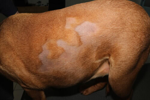
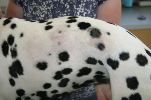
Canine recurrent flank alopecia (CRFA) is a relatively common dermatosis characterized by cyclic or seasonal non-inflammatory alopecia, preferentially localized to the flanks.
CRFA remains a condition whose etiopathogenesis is incompletely understood, and whose documentation in scientific literature, although existing, warrants an updated summary. This article aims to provide an update on current knowledge about CRFA, based on data from scientific publications. It will successively address epidemiology, etiopathogenic hypotheses, clinical presentation and variations, diagnostic approach including differential diagnoses and complementary examinations, typical histopathological characteristics, critical evaluation of proposed therapeutic options, as well as the prognosis for this condition.
Definition and Nomenclature
Several terms have been used to describe this clinical entity, including “seasonal flank alopecia,” “idiopathic cyclic flank alopecia,” or “cyclic follicular dysplasia.” However, none of these appellations are perfectly adequate. Hair loss is not always complete (sometimes simple changes in coat color or texture), it is not strictly confined to the flanks (other areas may be affected), and the seasonal or recurrent nature is not systematic in all affected individuals. The term “canine recurrent flank alopecia” is often preferred because it encompasses the most frequent localization and the cyclic nature often observed, while acknowledging these variations. The first formal description of this condition in the veterinary literature dates back to 1990 by Scott, who reported cases of fluctuating non-scarring alopecia in five spayed female dogs.
This condition, although visually striking to owners, is considered essentially cosmetic and is not associated with systemic signs or an alteration in the animal’s general health.
Appearance giving the impression that the dog has been clipped
Epidemiology
Breed Predispositions
CRFA is described in numerous canine breeds, but a clear breed predisposition is reported, strongly suggesting an underlying genetic component. This repeated observation in various studies implies that hereditary factors make certain lines more susceptible to developing the disease. The Boxer is the most frequently cited breed and appears particularly predisposed. Other breeds are also considered at increased risk, including the English Bulldog, Airedale Terrier, Schnauzer (miniature, standard, and giant), Bouvier des Flandres, Doberman, Labrador Retriever, Golden Retriever, Korthals Griffon, and Affenpinscher. More recent studies have also included the Rhodesian Ridgeback and Staffordshire Bull Terrier among affected breeds. An atypical form of recurrent flank alopecia has been specifically studied in the Cesky Fousek.
CRFA also exists in light-coated breeds
Age of Onset
The age of onset of CRFA is variable, ranging from 1 year to 11 years. However, the majority of cases develop the first clinical signs between 3 and 6 years of age. Specific studies on Boxers and Airedale Terriers have reported an average age of onset around 3.6 years, while another source mentions an average of 3.8 years. The average age at diagnosis is often around 4 years.
Sex and Reproductive Status Distribution
Initially, some publications reported an overrepresentation of spayed females. However, subsequent observations and larger studies have clearly established that there is no predisposition related to sex or reproductive status. Both intact and spayed males and females can be affected by CRFA.
Seasonal and Geographical Influence
One of the most characteristic aspects of CRFA is its frequent seasonality. In the Northern Hemisphere, alopecia typically appears during the months with the shortest daylight hours, generally between November and March or April. Regrowth then occurs spontaneously in the spring or summer.
Significantly, an inverse correlation has been observed in the Southern Hemisphere (Australia, New Zealand, Brazil), where the onset of alopecia also coincides with the short-day months (austral winter and spring). This observation is a strong argument in favor of the role of photoperiod as a major triggering factor. If classic seasonal environmental factors like temperature or humidity were paramount, one would expect onset during the same calendar season (e.g., winter) in both hemispheres, which is not the case. The systematic coincidence with decreasing day length (and thus increasing night length) strongly suggests the involvement of a biological mechanism sensitive to light cycles, likely mediated by the pineal gland and associated hormones like melatonin and prolactin.
However, it is important to note that this seasonality is not an absolute rule. Some dogs may present sporadic episodes, skip a season, or even develop alopecia that becomes permanent after several cycles.
Etiopathogenesis
Unknown Fundamental Cause
Despite racial predispositions and evocative seasonality, the exact etiology of CRFA remains unknown to date. Investigations conducted to identify an underlying systemic endocrine cause have been unsuccessful. Thyroid panels, adrenal function explorations (to rule out hyperadrenocorticism), and measurements of growth hormones or circulating sex hormones are generally within normal limits in dogs with CRFA. Nevertheless, the hypothesis of a localized alteration at the level of the hair follicles, such as a modification in the number or sensitivity of hormonal receptors, cannot be formally ruled out and remains a possible avenue.
Photoperiod Hypothesis
The influence of photoperiod (the daily duration of light exposure) is the etiopathogenic hypothesis best supported by clinical and epidemiological observations. The marked seasonality, correlated with the decrease in day length in both hemispheres, is the primary indicator. This hypothesis is reinforced by anecdotal reports describing dogs developing lesions outside the usual season when kept in dark or low-light environments. Conversely, attempts at prevention with phototherapy (exposure to intense artificial light for 15-16 hours a day during at-risk months) have shown some success in isolated cases, although controlled studies are lacking to confirm this approach.
Role of Melatonin
Melatonin is a neurohormone primarily synthesized by the pineal gland during darkness; its production is therefore inversely proportional to day length. It plays a fundamental role in regulating circadian and seasonal rhythms, including the hair cycle and seasonal molting, in many mammalian species. The main hypothesis regarding CRFA postulates that insufficient endogenous melatonin production, or an alteration in its reception or signaling at the hair follicles in genetically predisposed individuals, could be a key factor in pathogenesis. Melatonin could act directly on receptors present on follicular cells or indirectly by modulating the secretion of other hormones involved in the hair cycle, such as melanocyte-stimulating hormone (MSH) or prolactin. The empirical use of melatonin as a treatment is based on this hypothesis.
Role of Prolactin
Prolactin is another hormone whose secretion is influenced by photoperiod, often inversely to that of melatonin (an increase in melatonin tends to decrease prolactin levels). In some species like sheep, the decrease in prolactin induced by increased melatonin is associated with the induction of winter coats. Studies in mice and sheep have shown that prolactin can have inhibitory effects on the anagen (growth) phase of the hair follicle, potentially reducing hair length, shortening anagen, inducing shedding (exogen), or prolonging the telogen (resting) phase. Although no study has specifically evaluated the role of prolactin in CRFA, its involvement in the photoperiodic regulation of the hair cycle makes it a potential player in the pathogenesis of this condition.
Genetic Factors
The strong predisposition observed in certain breeds (Boxer, Airedale, English Bulldog, etc.) is a major argument in favor of a genetic component in CRFA. Genetic studies have begun to explore this avenue. A genome-wide association mapping study was conducted on an atypical form of CRFA (aCRFA) in the Cesky Fousek. This study identified several chromosomal regions (loci) associated with the disease, suggesting a polygenic basis (involving multiple genes) rather than a single mutation. The analysis highlighted 144 candidate genes potentially involved in four main metabolic pathways: collagen formation, muscle structure and contraction (potentially related to the arrector pili muscle), lipid metabolism (which can influence follicular development signaling pathways like WNT or SHH), and the immune system. Furthermore, genes related to circadian rhythm regulation and melatonin metabolism were among the candidates, reinforcing the link with photoperiod.
The demonstration of such diverse metabolic pathways (collagen, muscle, lipids, immunity) associated with aCRFA suggests unexpected complexity. Rather than a simple direct hormonal deregulation of the hair cycle, the pathogenesis of CRFA could involve a more fundamental disruption of skin homeostasis or the hair follicle’s very structure. These intrinsic structural or metabolic alterations, of genetic origin, could make the follicles abnormally sensitive to seasonal or hormonal triggers (photoperiod, melatonin, prolactin). This model goes beyond the simple causal chain “photoperiod → hormone → cycle arrest” and suggests a complex interaction between inherited tissue susceptibility and triggering environmental factors.
It should be noted that another study specifically targeting the MLPH gene (associated with color dilution) in Rhodesian Ridgebacks did not find an association with CRFA in this breed. This indicates that the genetic basis may be heterogeneous across breeds or that the genes involved are different from those related to pigmentation.
Follicular Mechanisms
Functionally, CRFA is considered an abnormality of the hair cycle, often described as “Hair Cycle Arrest.” Histopathology suggests a major defect in the initiation or progression of the anagen (growth) phase. This leads to an accumulation of hair follicles in the telogen (resting) phase or, after shedding of telogen hair, without immediate replacement.
The term “cyclic follicular dysplasia” is sometimes used, referring to the morphologically abnormal appearance of follicles observed histologically. Follicles may appear atrophic, deformed, with irregular basal structures. However, the term “dysplasia” (abnormal development) can be debated, with some preferring to focus on the functional aspect of cycle arrest.
Diagnosis
Clinical Approach
The suspected diagnosis of CRFA relies primarily on a detailed anamnesis and a rigorous clinical examination. Key anamnesis elements include belonging to a predisposed breed, typical age of onset (young adult to middle-aged adult), seasonal and recurrent nature of alopecia episodes (if present), and absence of general signs or pruritus.
Dermatological examination looks for cardinal clinical signs:
- Non-inflammatory and non-pruritic alopecia: This is an essential characteristic. The underlying skin is generally not red, thickened, or irritated, and the dog does not scratch.
- Typical location: The affected area predominates on the flanks (lateral or dorsolateral thoracolumbar region). It is often bilateral, but symmetry is not always perfect, and unilateral involvement, although rare, is possible.
- Appearance of lesions: Alopecic areas are generally well-demarcated, with sharp edges, sometimes irregular or serpiginous, forming “geographic map” patterns. The size of the lesions varies, from a few centimeters to almost the entire thoracolumbar region.
- Hyperpigmentation: Dark discoloration (black or brown) of the skin in alopecic areas is very frequent but not constant. Its absence does not rule out the diagnosis, as the ability to hyperpigment varies by breed and individual.
- Easy epilation: At the beginning of a shedding episode, hairs at the periphery of the lesions or in affected areas can often be easily epilated by gentle traction (positive trichogram).
It is also important to be aware of atypical presentations that may complicate the initial diagnosis: involvement of the muzzle or periorbital region (particularly in Labrador and Golden Retrievers), more generalized involvement, or simple color changes (e.g., aurotrichia – hair becoming golden) or texture changes of the flank coat without visible alopecia. Rare cases associated with histological interface dermatitis have also been described.
Differential Diagnoses
Given that symmetrical non-inflammatory alopecia is a presenting complaint that can correspond to several conditions, it is crucial to establish a rigorous differential diagnosis to rule out other potential causes. The main categories to consider are:
- Endocrinopathies: Hypothyroidism and hyperadrenocorticism (spontaneous or iatrogenic) are major differentials. Unlike CRFA, these diseases are often accompanied by systemic signs (lethargy, polyuria-polydipsia, weight gain, etc.) and other skin anomalies (thin skin, comedones, recurrent pyoderma). Sex hormone imbalances are rarer but possible.
- Other non-inflammatory alopecias: Alopecia X (sharing clinical and sometimes histological similarities), color dilution alopecia, various specific follicular dysplasias in certain breeds, telogen effluvium (massive shedding after stress) or anagen effluvium (shedding during the growth phase, rare), and “pattern baldness” should be considered.
- Infectious/parasitic causes: Although CRFA is non-inflammatory, generalized demodicosis or dermatophytosis can sometimes mimic symmetrical alopecia, especially early on. Bacterial folliculitis can also occur secondarily on alopecic areas of CRFA.
- Others: Sebaceous adenitis (inflammation of the sebaceous glands), post-clipping alopecia (regrowth failure after shaving), alopecia areata (immune mechanism), post-injection skin reactions (vaccine, medications), or traction alopecia (due to elastic bands, for example) are other possible differentials, although often with a different distribution or clinical history.
Additional Examinations
Confirmation of the CRFA diagnosis and exclusion of differentials require additional examinations:
- First-line examinations: Deep skin scrapings, trichograms (microscopic examination of hairs), skin cytology (search for bacteria, yeasts) and possibly fungal culture are indispensable to rule out demodicosis, dermatophytosis or secondary infection.
- Blood and hormonal tests: A hemogram and biochemical panel are useful to evaluate the general condition and look for signs of endocrinopathy. Specific hormonal assays, at least total T4 and TSH, are necessary to rule out hypothyroidism. In case of suspected hyperadrenocorticism, low-dose dexamethasone suppression or ACTH stimulation tests may be indicated.
- Skin biopsies: Histopathological examination of skin biopsies is considered the key step to confirm suspected CRFA and rule out other dermatoses with a similar presentation. It is recommended to take several biopsies (6 to 8 mm diameter punch) from within characteristic alopecic lesions. The timing of the biopsy is important, as histological lesions can vary depending on the stage of evolution (shedding phase, steady state phase, regrowth phase). Ideally, biopsies should be taken when alopecia is well established.
It is crucial to understand that, although histopathology provides very suggestive elements, the lesions observed in CRFA (infundibular hyperkeratosis, atrophic/dysplastic follicles) are not strictly pathognomonic and can sometimes be found, to varying degrees, in other conditions such as certain endocrinopathies or follicular dysplasias. Therefore, the definitive diagnosis of CRFA does not rely solely on biopsy but on an integrative approach. It requires the convergence of several elements: a compatible clinical presentation (breed, age, seasonality, appearance of lesions), rigorous exclusion of differential diagnoses (especially endocrine and parasitic) through appropriate examinations, and histopathological results compatible with non-inflammatory alopecia presenting the follicular characteristics described for CRFA.
Histopathology
General Characteristics
Histopathological examination of skin biopsies taken from alopecic areas in a dog with CRFA typically reveals non-inflammatory alopecia. Inflammatory infiltrate in the dermis or around the follicles is generally absent or very discreet, which is an important criterion for differentiating CRFA from alopecias of inflammatory or infectious origin.
Follicular Abnormalities
The most characteristic changes concern the hair follicles and their associated structures:
- Infundibular hyperkeratosis: This is one of the most constant and striking signs. It is a significant thickening of the cornified layer (keratin) at the upper portion of the hair follicle (infundibulum). This excess keratin can obstruct the follicular opening and sometimes extend to secondary follicles or sebaceous gland ducts.
- Infundibular dilation: The infundibula may appear significantly enlarged, forming cystic structures filled with keratin lamellae.
- Follicular atrophy and dysplasia: The middle and deep portions of the hair follicles (isthmus and bulb) are often atrophic, i.e., reduced in size. They may also show dysplastic features, with irregular, tortuous shapes, and poorly defined or deformed bulb structures.
- “Witch’s feet” or “octopus” appearance: These evocative terms describe a peculiar appearance of atrophic/dysplastic follicles, characterized by irregular and branching epithelial projections extending from the base of the follicle into the surrounding dermis. This appearance is considered very suggestive of CRFA, although not exclusive.
- Hair cycle arrest: Analysis of hair cycle phases reveals a clear predominance of follicles in the telogen (resting) or catagen (regressive transition) phase. Follicles in the anagen (active growth) phase are rare or absent in biopsies taken during the alopecic phase. An increase in the number of follicles in the kenogen phase (empty follicles after telogen hair shedding, before the start of a new anagen cycle) is also a frequent finding, indicating a defect in the induction of the new growth phase. It should be noted that the distribution of phases can be highly variable from one individual to another and depends heavily on the timing of the biopsy relative to the disease cycle. If the biopsy is performed late or during the regrowth phase, anagen follicles may be observed.
Other Findings
Other changes may be observed:
- Melanin aggregates: Melanin pigment clumps may be present in the lumen of dilated infundibula, within follicular epithelial cells, or even within associated sebaceous glands.
- Epidermis and Dermis: The epidermis is generally of normal thickness, sometimes discretely hyperplastic or superficially hyperpigmented. The dermis shows no significant abnormalities, particularly no inflammation or fibrosis.
Comparison with Other Alopecias
It is important to note that certain histological characteristics of CRFA are not exclusive. The increase in follicles in the kenogen phase is a common sign in several hair cycle disorders, indicating difficulty in initiating the anagen phase. Alopecia X, for example, also shows a strong predominance of follicles in the telogen phase and a low percentage of anagen, sometimes making distinction difficult based on histology alone. However, the marked dysplastic features, particularly the “witch’s foot” image, appear more characteristic of CRFA. The absence of marked inflammation helps differentiate CRFA from alopecias of infectious, parasitic, or immune origin (such as alopecia areata or sebaceous adenitis).
Prognosis
Evolution and Recurrences
CRFA is, by definition, an often recurrent condition. In many dogs, episodes of alopecia occur cyclically, typically annually, in connection with seasonal changes in photoperiod.
However, the evolution is marked by significant inter-individual variability:
- A significant proportion of dogs (estimated at around 20% in one source) will experience only one episode of CRFA in their lifetime, with no subsequent recurrence.
- Other dogs will exhibit very regular cycles, with hair loss occurring at the same time each year.
- Some individuals may “skip” a season, not presenting alopecia in a given year, only to recur the following year.
- Finally, in some dogs, particularly those who have experienced several recurrent cycles, the alopecia may eventually become permanent, or regrowth between episodes may become increasingly incomplete.
Hair Regrowth
Spontaneous hair regrowth is a frequent characteristic of CRFA. It generally occurs within 3 to 8 months after the onset of shedding, although longer delays, up to 14 or even 18 months, have been reported in some cases.
The quality of the regrowing coat is often altered. New hairs may have a different color (often darker, but sometimes lighter or golden – aurotrichia) and/or modified texture (duller, drier, coarser) compared to the surrounding normal coat. As a result, even after regrowth, the previously alopecic area often remains visually identifiable.
The prognosis regarding complete regrowth, with hair of identical quality and color to the original, is therefore variable and largely unpredictable for a given individual.
Impact on Quality of Life
It is essential to re-emphasize that CRFA is a strictly cosmetic condition. It does not affect the dog’s general health, well-being, or longevity in any way. The vital prognosis is excellent. The main impact is aesthetic for the owner.
This benign and often self-resolving nature fully justifies the conservative therapeutic approach (abstention or “benign neglect”) as a first-choice option. The absence of pain or associated systemic disease, combined with the high probability of spontaneous regrowth and the uncertain efficacy of treatments like melatonin (demonstrated in a controlled study), makes careful observation a medically sound strategy. It helps avoid potentially unnecessary interventions, costs, and constraints for a condition that does not impair the animal’s quality of life.
Conclusion
Canine recurrent flank alopecia (CRFA) is a dermatosis characterized by episodes of non-inflammatory alopecia, primarily affecting the flanks in a cyclic or seasonal manner. It preferentially affects certain breeds such as the Boxer, English Bulldog, and Airedale Terrier, suggesting a genetic predisposition. The exact etiopathogenesis remains unknown, but the influence of photoperiod is strongly suspected, potentially involving dysregulation in the production or signaling of melatonin and/or prolactin at the hair follicles. Recent genetic studies suggest a complex polygenic basis involving various metabolic pathways beyond simple hormonal regulation. Diagnosis relies on an evocative clinical picture (breed, seasonality, lesion appearance), rigorous exclusion of differential diagnoses (particularly endocrinopathies and parasites), and compatible histopathological examination (infundibular hyperkeratosis, atrophic/dysplastic follicles, absence of inflammation, hair cycle arrest). The vital prognosis is excellent, but complete hair regrowth and prevention of recurrence remain unpredictable.
Bibliography
- Vandenabeele S, Declercq J, De Cock H, Daminet S. Canine recurrent flank alopecia: a synthesis of theory and practice. Vlaams Diergeneeskd Tijdschr. 2014;83:275-81.
- Bassett RJ, Burton GG, Robson DC. Recurrent flank alopecia in a Tibetan Terrier. Aust Vet J. 2005 Jun;83(6):346-8.
- Verschuuren MUMY, Schlotter YM, van Geijlswijk IM, van der Lugt JJ, Gehring R. The efficacy of subcutaneous slow-release melatonin implants in the prevention of canine flank alopecia recurrence is uncertain: A double-blind, randomized, placebo-controlled study. Vet Dermatol. 2022 Dec;33(6):553-e111.
- Neradilová S, Schauer AM, Hayward JJ, Brunner MAT, Bohutínská M, Jagannathan V, Connell LB, Boyko AR, Welle MM, Černá Bolfíková B. Genomic and Transcriptomic Characterization of Atypical Recurrent Flank Alopecia in the Cesky Fousek Dog Breed Points to Novel Candidate Genes and Pathways. Genes (Basel). 2022 Apr 7;13(4):650.
- (Missing from extraction)
- Miller MA, Dunstan RW. Seasonal flank alopecia in boxers and Airedale terriers: 24 cases (1985-1992). J Am Vet Med Assoc. 1993 Dec 1;203(11):1567-72.
- Müntener T, Schuepbach-Regula G, Frank L, Rüfenacht S, Welle MM. Canine noninflammatory alopecia: a comprehensive evaluation of common and distinguishing histological characteristics. Vet Dermatol. 2012 Jun;23(3):206-e42.
- Lee JB, Kim JW, Park JK, Lee NS, Kim HJ, Yu DH, Kim SJ, Kim GT, Hwang CY. Seasonal Flank Alopecia in a Dog. J Vet Clin. 2014; 31(4): 341-343.
- Driemeier D, Fernandes CG, Wouters ATB, Wouters F. Canine cyclical flank alopecia in Southern Brazil. Braz J Vet Pathol. 2008; 1(2): 60-63.
- Verschuuren MUMY, Schlotter YM, Leegwater PAJ. Investigation of the association of the MLPH gene with seasonal canine flank alopecia in Rhodesian Ridgeback dogs. Canine Med Genet. 2024 Jan 18;11(1):1.
- Grant D. Canine flank alopecia. Veterinary Practice. 2016 Jul 1.
Related Searches
canine flank alopecia, rhodesian ridgeback, alopecia, flanks, dog, in dogs, dysplasia, hair, component, loss, hair loss, cases, condition, crfa, boxer, consultation, cycle, disorders, animal, shaving, pattern, growth, dermatosis, area, onset, clinical examination, hyperpigmentation, follicles, skin, disease, affected, breeds, map-like, bouvier des flandres, shedding, region, hypothyroidism, basis, pathology, episodes, reason, hypercorticism, health, weight gain, color, problem, formation, cushing’s syndrome, follicular dysplasia, approach, anomaly, coat, patterns, examination, etiopathogenesis, hypotheses, example, absence, duration, factors, knowledge, diagnosis, article, light, exposure, report, prognosis, differential diagnosis, character, molt, adult, studies, lesions

