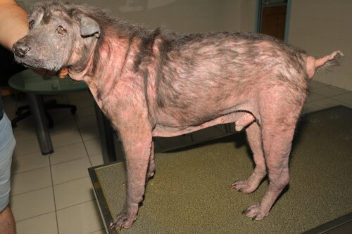
Canine atopic dermatitis (CAD) represents a major therapeutic challenge in veterinary dermatology, often requiring long-term management. Intralymphatic immunotherapy (ILIT) has progressively emerged as a promising alternative to conventional treatments, offering encouraging results in both human and veterinary medicine.
This innovative technique relies on the direct injection of allergens into the lymph nodes, allowing for faster and more effective desensitization than with conventional subcutaneous immunotherapy.

Severe Atopic Dermatitis in a Sharpei
The precision of the injection is a crucial parameter for successful treatment, particularly due to the allergen doses used, which are approximately 100 times lower than those in subcutaneous immunotherapy. Initially performed exclusively under ultrasound guidance, ILIT is now frequently practiced by simple palpation in many veterinary clinics. This evolution in practices raises legitimate questions about the potential impact of the injection method on therapeutic efficacy.
Materials and Methods
Study Population
This retrospective study focused on a substantial cohort of 109 dogs with canine atopic dermatitis, treated with ILIT between 2014 and 2022. The diagnosis of atopic dermatitis was based on rigorously validated clinical criteria, with a systematic prior exclusion of food allergy. Only dogs presenting non-seasonal pruritus were included in the study, to ensure the homogeneity of the study population and the reliability of the results.
Therapeutic Protocol
The immunotherapy protocol was standardized for all patients. The allergenic solutions used, supplied by a specialized laboratory (Heska AG, Fribourg, Switzerland), contained up to seven aqueous allergens, with an average of three allergens per preparation. Allergen selection was personalized for each patient, based on their specific sensitization profile and detailed clinical history. The final preparation consisted of an equal mixture of allergenic solution and aluminum hydroxide (Alhydrogel 2%, InvivoGen), administered at 0.2 mL every four weeks into a popliteal lymph node.
Injection Methodology and Evaluation
Two injection techniques were compared in this study: ultrasound-guided injection (U-ILIT), performed by a certified radiologist, and palpation-guided injection (P-ILIT), performed by a certified dermatologist. Patients received between three and six injections, according to a standardized protocol.
The evaluation of therapeutic efficacy was based on an in-depth comparative analysis of clinical status and symptomatic treatment needs between the beginning and the end of the injection protocol. A case was considered a responder when two essential criteria were simultaneously met: the possibility of reducing symptomatic treatments (oclacitinib, topical or systemic glucocorticoids) and the observation of significant clinical improvement, jointly validated by the owner and the treating veterinarian.
Results
Demographic Characteristics
The studied population showed great racial diversity, reflecting the clinical reality of canine atopic dermatitis. French Bulldogs represented the most frequent breed with fifteen individuals, followed by mixed-breed dogs (twelve individuals), West Highland White Terriers (nine individuals), and Labrador Retrievers (seven individuals). The average age of the patients was 3.5 years, with a distribution ranging from 1 to 10 years. Gender distribution was balanced, including fifty-nine males and fifty females.
Comparative Analysis of Injection Techniques
The results revealed a significant difference in efficacy between the two injection techniques. In the U-ILIT group, comprising 84 dogs, the positive response rate reached 60.7% (51 cases), while in the P-ILIT group, consisting of 25 dogs, only 28% of patients (7 cases) showed satisfactory improvement. This difference proved to be statistically significant (p=0.005), highlighting the potential importance of ultrasound guidance in the success of the treatment.
Further statistical analysis revealed no significant influence of other potentially confounding factors. The number of injections (p=0.08), the weight of the animals (p=0.36), and the breed of the patients (p>0.25 for the most represented breeds) showed no significant impact on the treatment response rate.
Discussion
This study highlights the crucial importance of injection precision in the efficacy of intralymphatic immunotherapy. The significantly higher response rate obtained with ultrasound guidance suggests that the technical quality of the injection is a determining factor for therapeutic success.
Nevertheless, several methodological limitations must be considered in the interpretation of these results. The retrospective nature of the study and the absence of standardized objective clinical scores like CADESI or PVAS constitute potential biases. The numerical imbalance between the groups (84 versus 25 patients) could also influence statistical interpretation, although the observed significance suggests a real difference in efficacy. Furthermore, the relatively short follow-up period does not allow for long-term evaluation of the observed therapeutic effects.
Conclusion
The results of this retrospective study strongly suggest that ultrasound guidance improves the efficacy of intralymphatic immunotherapy in the treatment of canine atopic dermatitis. This observation emphasizes the importance of technical precision in treatment administration and questions the relevance of an excessive simplification of injection protocols. These preliminary data nevertheless require confirmation by controlled prospective studies, which will also allow for evaluation of the impact of additional factors such as practitioner experience or optimal treatment duration.
FAQ
- Why prefer intralymphatic immunotherapy over conventional subcutaneous immunotherapy? ILIT allows for faster desensitization with significantly reduced allergen doses, potentially offering a better benefit-risk ratio and optimized therapeutic adherence.
- Is ultrasound guidance systematically indispensable for all patients? The results of this study suggest a significant benefit of ultrasound guidance, but further studies are needed to identify potential subgroups of patients for whom palpation might suffice.
- Does the injection technique influence the necessary treatment duration? This specific question was not directly explored in this study and would merit dedicated investigation, particularly to evaluate the potential impact of injection precision on the speed of the therapeutic response.
- Can practitioner experience compensate for the lack of ultrasound guidance? This variable was not specifically evaluated in this study but could be the subject of future research, particularly relevant for optimizing practitioner training.
- Are there patient selection criteria that can optimize ILIT results? This study did not identify clear predictive factors for treatment response, suggesting that the injection technique remains the main parameter determining therapeutic efficacy.
Fischer NM, Favrot C, Martini F, Rostaher A. Intralymphatic Immunotherapy with Ultrasound Guidance Seems to Be Associated with Improved Clinical Effect in Canine Atopic Dermatitis—A Retrospective Study of 109 Cases. Animals (Basel). 2024 Oct 11;14(20):2921. doi: 10.3390/ani14202921.
Related Searches
canine atopic dermatitis, dog, atopic in dogs, in dogs, blood test, genetic disease, symptom, itching, owners, article, immune system, life, options, animal, contact, mites, health, symptoms, points, licking, allergies, in dogs, pyoderma, types, atopic dogs, effect, necessity, eczema, barrier, ears, grasses, origin, fleas, skin barrier, predisposition, shar pei, flare-ups, pollen, causes, dermatosis, fatty acids, age, bites, inflammation, golden retriever, paws, side effects, crisis, collaboration, redness, antibodies, site, reaction, diseases, signs, skin, malassezia pachydermatis, cause