Feline eosinophilic granuloma complex is one of the three major dermatological syndromes observed in cats, along with miliary dermatitis and self-induced alopecia.
Author: William Bordeau
VetDerm Clinic
1 avenue Foch 94700 MAISONS-ALFORT
The more time passes, the more the term feline eosinophilic granuloma complex (EGC) and its various components appear unsuitable. Increasingly, it is suggested to avoid talking about eosinophilic plaques, eosinophilic granuloma, and indolent ulcer. Indeed, these are associations of clinical and histological terms which, moreover, do not fully reflect reality.
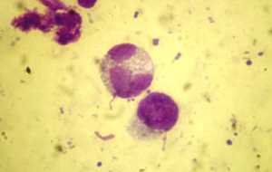
Eosinophils are among the poorly studied cells, both in companion animals and in humans. Initially, the hypothesis was that they only intervened in the body’s immune defenses against parasites, particularly helminths. Gradually, their importance in certain dermatoses, including allergic dermatitis, was discovered. Initially, eosinophils are found in the bloodstream. Then, through chemotaxis, they cross the vessels to reach the inflammatory site where they are then activated. This activation induces the release of numerous proinflammatory and immunomodulatory molecules. They also lead to the release, particularly through degranulation, of cytotoxic molecules that can act on helminths, bacteria, viruses, and tumor cells.
Photo 1: Cat Eosinophil
Feline eosinophilic granuloma complex is similar to Well’s syndrome, observed in humans. This is notably because bites from certain arthropods, as well as the presence of some fungi and the ingestion of certain medications, can lead to the appearance of this syndrome in humans. However, as in cats, the etiopathogenesis of Well’s syndrome is not fully known. It manifests as eosinophilic plaques primarily located at the extremities. Lesions can disappear spontaneously. During the active phase of the dermatosis, hypereosinophilia is noted.
The observation of a plaque, eosinophilic granuloma, or indolent ulcer on an animal simply qualifies the presence of an eosinophilic granuloma complex, but in no way allows for diagnosis. Indeed, this condition can be secondary to numerous causes. Thus, it is only the manifestation of a reactive process in response to various stimuli. It can be due to allergic dermatitis to arthropods, a food allergen, or an aeroallergen, but can also be secondary to an infectious dermatosis or a chemical cause. This eosinophilic granuloma complex can also be congenital. However, in many cases, it is idiopathic. But this does not mean that the search for an underlying cause should necessarily be neglected. There would also be a genetic predisposition, which would explain in particular that even if many cats have allergic dermatitis, relatively few are affected by an eosinophilic granuloma complex.
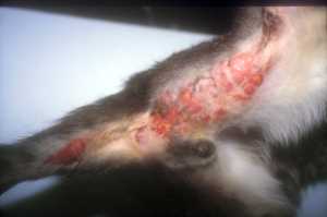
This complex can manifest as a plaque or a granuloma. These are erythematous, firm, and alopecic lesions. They can be observed in many parts of the body. Granulomas are generally linear and usually located on the caudal aspect of the thighs. There is also a buccal variant and a chin variant, which are non-linear. Granuloma appears more often in young cats, and plaque in adult animals.
Photo 2: Eosinophilic granuloma
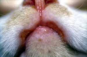
The eosinophilic granuloma complex can also manifest as a shiny, non-bleeding ulcer with a color ranging from yellow to brown. It is observed on the upper lip and often starts opposite a canine tooth. It can be unilateral or bilateral and symmetrical. This ulcer is not painful or pruritic, unlike plaques and granulomas which can cause sometimes intense pruritus. When present, erosions are also observed on these lesions. Furthermore, it is possible for plaques and granulomas to coexist on the same animal.
Photo 3: Indolent ulcer
These lesions can persist, but also disappear spontaneously. This simply reflects the lack of knowledge of this syndrome and the plurality of underlying causes.
There is currently a controversy over whether or not to include miliary dermatitis and allergic dermatitis due to mosquito bites in the eosinophilic granuloma complex, as there are histopathological similarities.
Identifying an underlying cause is a necessity. First, it is important to confirm the eosinophilic granuloma complex. A skin impression smear is thus performed on the lesions to demonstrate the presence of eosinophils. It is also possible to observe degenerated neutrophils and intracellular or extracellular bacteria in varying numbers. In case of doubt, skin biopsies are performed. In parallel, it is essential to rule out all dermatoses included in the differential diagnosis. While it is extremely broad, it is especially important to consider the form of the eosinophilic granuloma complex (plaque, granuloma, or ulcer) and the location of the lesions.


