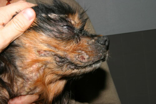
Psoriasiform lichenoid dermatosis constitutes an unusual cutaneous manifestation in dogs, closely associated with the administration of calcineurin inhibitors and complicated by staphylococcal infection. This dermatological condition, characterized by distinctive hyperkeratotic lesions, raises fundamental questions concerning the complex interactions between therapeutic immunomodulation and cutaneous pathogens.
The retrospective analysis of twenty-eight canine cases over a period extending from 2015 to 2023 provides insight into the clinical, histopathological and therapeutic features of this poorly understood pathological entity, while exploring the genomic characteristics of staphylococcal strains involved in its genesis.
Epidemiological characteristics and clinical presentation
Examination of the study population reveals a remarkable absence of breed or sex predisposition. The cohort comprises fifteen purebred dogs and eight mixed-breed individuals, with notable representation of three American Pit Bull Terriers, three Labrador Retrievers, two Boxers and two Greyhounds. The median age at onset is seven years, with variations ranging between three and twelve years, suggesting a predominance in adult animals.
The anatomical distribution of lesions presents substantial variability. Four cases manifest a single focal involvement, while seventeen present multifocal lesions and seven develop a regional or generalized form. Clinical manifestations are primarily characterized by hyperkeratotic to nodular plaques, frequently described as presenting a frond-like appearance. These cutaneous proliferations are generally accompanied by alopecia and affect various body regions including the head, neck, trunk and limbs. Seven animals manifest concomitant pruritus at the time of biopsy sampling.
Lichenoid dermatosis in a dog
Exposure to calcineurin inhibitors and associated factors
Twenty-seven of the twenty-eight documented dogs received a calcineurin inhibitor prior to lesional emergence. The median delay preceding the appearance of cutaneous manifestations is six months, with extremes varying from one to twenty-four months. This variable latency suggests differential individual susceptibility or the intervention of pathogenic cofactors.
Modified cyclosporine represents the predominant inhibitor, administered to twenty-three individuals either alone or in combination with ketoconazole. The distribution of pharmaceutical formulations is as follows: twelve dogs receive a generic microemulsified cyclosporine, seven benefit from a branded commercial preparation, one animal is treated with a compounded formulation, while three cases involve unspecified preparations. Seven patients receive cyclosporine and ketoconazole simultaneously, the latter being used for its cytochrome P450 3A inhibition properties, allowing an increase in blood cyclosporine concentrations.
Dosage analysis reveals that a significant proportion of twelve dogs receive doses exceeding established recommendations, with a median of 10 mg/kg/day and maximum values reaching 15.2 mg/kg/day. For animals treated exclusively with cyclosporine, the median dose amounts to 7 mg/kg/day. Patients receiving the cyclosporine-ketoconazole combination benefit from a reduced cyclosporine dosage, with a median of 2.7 mg/kg/day, compensated by concomitant administration of ketoconazole at a median dose of 4.9 mg/kg/day.
Topical tacrolimus constitutes a less frequent therapeutic modality, concerning five animals. Three of them receive ophthalmic preparations for the treatment of keratoconjunctivitis sicca, while two benefit from cutaneous applications. All dogs treated with topical tacrolimus develop lesions at or in immediate proximity to the application site, suggesting a direct causal relationship.
Underlying conditions and concomitant therapeutics
Therapeutic indications justifying the administration of calcineurin inhibitors prove diverse. Six animals present refractory atopic or allergic dermatitis, three suffer from protein-losing enteropathy, two manifest respectively immune-mediated encephalitis or myelitis, pemphigus foliaceus, inflammatory bowel disease, immune thrombocytopenia or immune-mediated polyarthritis. Isolated cases concern sterile pyogranulomatous dermatitis, protein-losing nephropathy, pure red cell aplasia and immune blepharitis.
Sixteen patients receive other immunomodulatory agents simultaneously. Six animals benefit from prednisone, two receive lokivetmab injections, two are treated with oclacitinib, two with budesonide, while individual cases concern the administration of ophthalmic solutions combining neomycin, polymyxin B and dexamethasone, ophthalmic prednisolone acetate or levothyroxine. This immunomodulatory polytherapy could potentiate alterations in the cutaneous immune response.
Cytological and microbiological investigations
Lesional cytological examination, performed in twenty-six animals, identifies bacterial cocci in twenty-two cases. Three of these samples also reveal the presence of bacilli and yeasts compatible with Malassezia spp. Four samples present no identifiable bacteria. This high prevalence of superficial bacterial colonization supports the hypothesis of a determining pathogenic role of infection in lesional genesis.
Aerobic cultures, performed on nine samples among cytologically positive cases, isolate exclusively Staphylococcus pseudintermedius. Eight cultures reveal this species as the sole pathogen, while one sample additionally presents slight growth of group D Enterococcus sp. and Escherichia coli. This microbiological uniformity suggests a specific relationship between S. pseudintermedius and the development of psoriasiform lichenoid dermatosis.
Histopathological characteristics
Microscopic examination reveals a remarkably constant histological pattern. All samples present a lymphoplasmacytic lichenoid band in the superficial dermis. The epidermis manifests acanthosis with formation of projections in the form of epithelial ridges, conferring the psoriasiform aspect. Bacterial cocci are invariably observed in crusts or microabscesses. In three individuals, the lichenoid lymphoplasmacytic infiltrate extends periadnexally to the level of the follicular isthmus.
Parakeratotic hyperkeratosis characterizes the stratum corneum, associated with orthokeratosis in ten cases. Follicular hyperkeratosis is occasionally observed in six samples. These epidermal architectural modifications, associated with specific dermal inflammation, allow histopathological distinction from other proliferative cutaneous conditions such as zinc-responsive dermatosis, viral papillomas, pigmented viral plaques or proliferative lymphocytic mural folliculitis of Labrador Retrievers.
Therapeutic modalities and clinical evolution
Therapeutic management generally combines antimicrobials and modification of the calcineurin inhibitor protocol. Fourteen animals receive antimicrobials prior to diagnosis, while twenty-two benefit from post-diagnostic antibiotic therapy. Three patients are treated exclusively with antiseptics or topical antimicrobials. Therapeutic classes employed include doxycycline, clindamycin, rifampin, cephalosporins, terbinafine, marbofloxacin, tylosin, ciprofloxacin, sulfadimethoxine-ormetoprim, amoxicillin and minocycline.
Adjustment of the calcineurin inhibitor is performed according to various modalities: thirteen cases involve complete discontinuation, ten a dosage or frequency reduction, while one protocol remains unchanged. For four animals, information concerning this therapeutic modification remains unavailable. Two particular cases concern discontinuation of ketoconazole to effectively reduce cyclosporine exposure, four involve dosage reduction, and three a reduction in administration frequency.
Evaluation of therapeutic results reveals that four dogs obtain improvement greater than fifty percent, while eighteen achieve complete lesional resolution after antibiotic therapy and modification of calcineurin inhibitor treatment. Two animals manifest no improvement despite antibiotic therapy, these cases corresponding to situations where cyclosporine was never reduced or whose therapeutic adjustment was delayed by owner non-compliance. Six patients are lost to follow-up, making evaluation of their final lesional status impossible.
Recurrences and long-term remission
Three animals having initially obtained complete resolution develop recurrence. The first case, on cyclosporine and ketoconazole, presents lesional reappearance twelve months after antimicrobial discontinuation. A new series of biopsies excludes a differential diagnosis of viral papillomas, and additional systemic antibiotic therapy leads to secondary complete resolution. The second patient, treated empirically with partial improvement, manifests worsening three months after initial identification. Cyclosporine discontinuation following histopathological confirmation allows lasting remission. The third case, in which only ketoconazole is discontinued while cyclosporine continues, obtains resolution under clindamycin but recurs three months post-treatment. Isolation of methicillin-resistant S. pseudintermedius motivates institution of topical antiseptic therapy, leading to resolution in thirty days.
At the time of manuscript submission, nineteen animals presenting substantial improvement or complete resolution remain in clinical remission according to available information. This favorable proportion suggests that early diagnostic recognition and appropriate therapeutic adaptation generally allow effective control of the condition.
Pathogenic and clinical implications
The near-universal presence of bacteria in lesions, combined with the therapeutic efficacy of antimicrobials, solidly supports the hypothesis according to which staphylococcal infection constitutes a central pathogenic element. This dermatosis would thus represent an atypical immune reaction to bacterial infection, facilitated by immunomodulation induced by calcineurin inhibitors. The conjunction of calcineurin inhibition and exposure to staphylococcal strains carrying specific virulence factors could alter the normal cutaneous immune response, leading to the distinctive histopathological pattern observed.
The observation that several animals present dosages exceeding established recommendations suggests a possible dose-dependent relationship in lesional development. This hypothesis would merit confirmation by prospective controlled studies. The single documented case of therapeutic monitoring reveals a serum concentration largely exceeding the target therapeutic interval, reinforcing the plausibility of a dose-dependent effect.
The finding of lesional association with topical tacrolimus, particularly at high ophthalmic concentrations of one percent, extends the spectrum of calcineurin inhibitors involved beyond oral cyclosporine. The development of facial lesions in an animal receiving only ophthalmic tacrolimus suggests significant systemic absorption of this compound.
Differential diagnostic considerations
Histopathological distinction between psoriasiform lichenoid dermatosis and other acanthotic hyperkeratotic conditions rests on specific criteria. Zinc-responsive dermatosis classically presents severe parakeratosis extending into follicular infundibula with spiral formation, contrasting with the serocellular crusts and discrete intraepidermal pustules characterizing the present condition. Regressing viral papillomas, in the absence of inclusion bodies or viral cytopathic effect, manifest cytotoxic interface dermatitis rather than the lichenoid inflammatory band observed here. Proliferative lymphocytic mural folliculitis of Labrador Retrievers is distinguished by follicular acanthosis and hyperplasia accompanied by keratinocyte apoptosis and mild mural cytotoxic interface folliculitis.
Clinically, differentiation from classic bacterial folliculitis is required, the latter typically manifesting as papules and pustules rather than hyperkeratotic plaques. Recognition of this distinctive lesional pattern, associated with history of calcineurin inhibitor exposure and cytological identification of abundant cocci, strongly orients toward presumptive diagnosis, confirmed subsequently by histopathological analysis.
The entirety of these observations defines psoriasiform lichenoid dermatosis as a well-characterized clinicopathological entity, occurring in adult dogs regardless of breed, closely linked to the administration of calcineurin inhibitors in various formulations and intimately associated with S. pseudintermedius infection. Early diagnostic recognition allows institution of appropriate antimicrobial treatment and therapeutic adjustment of calcineurin inhibitors, generally leading to favorable clinical outcomes with substantial or complete lesional resolution in the majority of documented cases.
Davis ER, Mauldin EA, Cain CL, Cole S, Bradley CW. Clinical features, treatment and outcomes of dogs with psoriasiform lichenoid dermatosis associated with calcineurin inhibitor therapy. Vet Dermatol. 2025;0:1-14.
