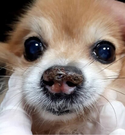
Pemphigus foliaceus represents a rare but significant autoimmune bullous dermatosis in dogs. Systemic corticosteroids are usually the first-line treatment, but their variable efficacy and significant side effects justify the exploration of therapeutic alternatives, as was the case in this clinical case.
Pathophysiological Context
Pemphigus foliaceus is the most common autoimmune dermatosis in dogs and cats. Its physiopathological mechanism relies on the production of autoantibodies directed against epidermal cells. This immune reaction causes a rupture of desmosomal junctions in the epidermis, leading to a phenomenon of acantholysis. The acantholytic cells thus formed constitute a characteristic cytological marker of this condition.
The clinical manifestations of pemphigus foliaceus are initially characterized by the appearance of superficial pustules that typically evolve into the formation of crusts, epidermal collarettes, and erosions. Alopecia and hyperkeratosis may also be observed in the clinical picture. The preferentially affected anatomical regions include the head, face, and ears, with the possibility of nasal depigmentation.
Diagnostic Approach
The diagnosis of pemphigus foliaceus is based on a methodical approach combining several complementary elements. A detailed anamnesis and clinical examination constitute the essential first steps of this process. The observation of intact pustules represents a particularly relevant semiological element to guide the diagnosis.
Cytopathological examination of the pustules generally reveals the presence of acantholytic cells, neutrophils, and sometimes eosinophils. However, definitive confirmation of the diagnosis requires histopathological analysis of skin biopsies. Typical histological features include subcorneal pustules, acantholytic cells, and an inflammatory infiltrate composed mainly of neutrophils, sometimes accompanied by eosinophils.
Conventional Therapy and Its Limitations
The reference treatment for pemphigus foliaceus traditionally relies on the administration of prednisolone. This molecule exerts a powerful immunosuppressive action, aiming to control the production of autoantibodies and reduce skin inflammation.
However, the efficacy of this conventional therapy shows considerable variability among individuals. Zhou et al. (2021) reported an average remission time of 56 days with corticosteroid therapy. More problematic, prolonged use of corticosteroids is frequently accompanied by significant adverse effects, including diabetes, muscle atrophy, muscle weakness, and calcinosis cutis.
Oclacitinib: Mechanism of Action and Therapeutic Applications
Oclacitinib stands out as a Janus kinase (JAK) inhibitor initially developed for the treatment of canine allergic diseases, particularly atopic dermatitis.
This molecule offers several interesting pharmacological advantages, including rapid action, demonstrated clinical efficacy in allergic dermatoses, and a favorable safety profile even with prolonged use. These characteristics have led to exploring its therapeutic potential in other dermatological contexts, including autoimmune skin diseases.
In humans, JAK inhibitors have shown promising results in the treatment of various autoimmune pathologies. This observation is particularly relevant in the context of human pemphigus vulgaris, where an overexpression of JAK3 enzymes has been demonstrated in skin lesions compared to healthy skin. This correlation could explain the potential efficacy of JAK inhibitors in autoimmune dermatoses.
In veterinary medicine, several clinical cases have documented the successful off-label use of oclacitinib in autoimmune skin conditions. Aymeric and Bensignor (2017) reported the efficacy of this molecule in a German Shepherd dog with subepidermal bullous dermatosis. Significant improvement in clinical signs was observed after one month of treatment, with no notable adverse effects even after 12 months of use.
Similarly, Carrasco et al. (2021) documented the off-label efficacy of oclacitinib in a cat with pemphigus foliaceus. A decrease in pruritus and the severity of skin lesions was noted from the first week of treatment.
Clinical Case Presentation
This study reports the case of an 11-year-old male German Spitz dog, presented for consultation due to nasal depigmentation. The anamnesis revealed a previous treatment with daily oral cyclosporine and prednisolone every other day, with no satisfactory clinical response.
Clinical examination revealed an alteration in the architecture of the nasal planum, characterized by the presence of crusts, depigmentation, and ulcerations. Blood tests revealed thrombocytopenia, leukocytosis, lymphocytosis, and eosinophilia. Serum biochemical parameters (alanine aminotransferase, alkaline phosphatase, urea, creatinine, triglycerides, cholesterol, total proteins and fractions) were within reference ranges.
A previous histopathological examination had been inconclusive. Complementary tests included a negative serology for leishmaniasis, a negative 4DX test, and a cytopathological examination with no significant abnormalities. A discontinuation of prednisolone treatment for 20 days was recommended to perform a new skin biopsy.
Histopathological analysis of this second sample revealed orthokeratotic hyperkeratosis associated with subcorneal pustules containing segmented neutrophils and discrete aggregates of acantholytic cells. These characteristics were compatible with a diagnosis of pemphigus foliaceus. Extensive foci of necrosis and epidermal ulcerations were also observed in some sections.
Therapeutic Approach with Oclacitinib
Considering the lack of clinical response to previous treatments (oral corticosteroids and cyclosporine), oral oclacitinib therapy was initiated for 14 days.
After this initial treatment period, significant improvement in nasal lesions was observed, with healing of ulcers and progressive normalization of platelet count. Given this favorable outcome, the frequency of administration was reduced to once daily.
After 30 days of treatment, complete resolution of nasal lesions was observed, with no persistent depigmentation. Regular clinical and biological monitoring was recommended. Notably, no adverse effects were reported throughout the treatment period.
Discussion and Perspectives
This clinical case illustrates the potential efficacy of oclacitinib in the management of canine pemphigus foliaceus refractory to conventional treatments. Several aspects deserve to be highlighted and analyzed.
Pemphigus foliaceus, like any autoimmune disease, typically involves prolonged, even lifelong, treatment. In this context, the adverse effects associated with long-term corticosteroid therapy constitute a major concern in animals. Oclacitinib, characterized by a low incidence of side effects even with prolonged use, could represent a particularly interesting therapeutic alternative.
The precise mechanism of action of oclacitinib in animal autoimmune diseases has not yet been fully elucidated. Nevertheless, by analogy with data from human medicine, the inhibition of JAK enzymes could interfere with the signaling of cytokines involved in the pathogenesis of pemphigus foliaceus.
The case reported here demonstrates satisfactory therapeutic efficacy. Unlike other cases described in the literature, a reduction in the frequency of administration to once daily proved sufficient to maintain clinical remission. This observation suggests the possibility of maintenance treatment at a reduced dose, potentially limiting the risks of long-term adverse effects.
The absence of notable side effects in this case corroborates the available data in the literature regarding the safety of oclacitinib. This characteristic is particularly advantageous for the treatment of chronic diseases requiring prolonged therapy.
These promising results join other similar clinical observations, notably the case of a dog with cutaneous lupus erythematosus successfully treated with oclacitinib, initially twice daily for 15 days, then once daily (Lima & Cunha, 2022).
Oclacitinib could represent a particularly interesting therapeutic option for patients for whom corticosteroid therapy is contraindicated, such as diabetic dogs or those with hyperadrenocorticism. Its corticosteroid-sparing profile also constitutes a notable advantage, as recently demonstrated (Hernandez-Bures et al., 2023).
Conclusion
Oral administration of oclacitinib proved effective and safe in the treatment of pemphigus foliaceus in a dog refractory to conventional therapy with corticosteroids and cyclosporine. This molecule could constitute a new therapeutic option for this autoimmune condition.
The complete resolution of skin lesions, the absence of adverse effects, and the possibility of maintenance treatment at a reduced dose represent significant advantages compared to classic therapeutic approaches.
However, further studies involving a larger number of patients are needed to evaluate the long-term efficacy of oclacitinib in canine pemphigus foliaceus, determine optimal dosages, and identify potential predictive factors for treatment response.
Silva MMC, Bernardini M, Lopes NL. Use of Oclacitinib in the treatment of pemphigus foliaceus in a dog: case report. Braz J Vet Med. 2025;47:e009024. DOI: 10.29374/2527-2179.bjvm009024.
