Feline immune-mediated skin diseases comprise a complex set of rare but potentially serious conditions that can mimic various infectious or allergic dermatoses. This second part of a series on immune-mediated dermatoses examines six distinct pathological entities characterized by specific pathophysiological mechanisms.
Early identification of these conditions is crucial given their potentially fatal progression and the systemic repercussions they can cause. A thorough understanding of their clinical presentation, histopathological characteristics, and therapeutic modalities is an essential prerequisite for optimizing the management of these particularly fragile patients.
Erythema Multiforme and Toxic Epidermal Necrolysis Syndrome
Erythema Multiforme: Mechanisms and Manifestations
Erythema multiforme represents an acute immune reaction affecting the skin and mucous membranes, whose pathogenesis involves the activation of cytotoxic T lymphocytes directed against antigen-loaded keratinocytes. This activation triggers an inflammatory cascade characterized by epidermal cell lysis and keratinocyte apoptosis. Classification distinguishes two forms based on the extent of mucosal involvement and the presence of systemic signs: the minor form, limited to cutaneous manifestations, and the major form, accompanied by mucosal involvement and general symptoms.
Triggers identified in felines primarily include drugs, although upper respiratory viral infections and certain vaccinations have been reported. An association with feline herpesvirus type 1 has been proposed in two cases presenting generalized exfoliative dermatitis with erosions and scales, accompanied by a history of recurrent respiratory infections. This hypothesis is supported by the detection of viral DNA in skin biopsies, suggesting a mechanism similar to that observed in humans with herpes simplex virus.
Clinical presentation varies considerably, ranging from localized maculopapular lesions with characteristic cockade formation on the ventral part of the body, to more extensive manifestations including generalized scales, alopecia, erosions, and ulcerations with or without mucocutaneous involvement. Unlike Stevens-Johnson syndrome and toxic epidermal necrolysis, erythema multiforme does not cause extensive epidermal detachment.
Stevens-Johnson Syndrome and Toxic Epidermal Necrolysis
These two entities constitute manifestations of the same pathological spectrum, differentiated only by the extent of epidermal detachment. Stevens-Johnson syndrome affects less than 10% of the body surface, the intermediate form (SJS/TEN overlap) affects 10 to 30% of the surface, while toxic epidermal necrolysis involves more than 30% of the epithelium.
The pathogenesis is based on a hypersensitivity reaction mediated by cytotoxic T lymphocytes, primarily triggered by drug exposure. In felines, beta-lactam antibiotics, organophosphorus insecticides, and d-limonene show a strong causal association. The ALDEN algorithm (Algorithm of Drug Causality for Epidermal Necrolysis), recently validated in humans, has been successfully used for evaluating drug causality in a recent feline case.
Clinical manifestations begin with painful and irregular erythematous macules and plaques, evolving into bullae formation and then into confluent epidermal detachment. Involvement of mucocutaneous junctions, oral, rectal, and conjunctival mucous membranes, as well as paw pads, is a frequent characteristic. In severe cases, necrolysis can extend to the respiratory and gastrointestinal epithelia, leading to major systemic complications including bronchial obstruction, profuse diarrhea, and multi-organ failure.
Plasmacytic Pododermatitis
Distinctive Clinical Features
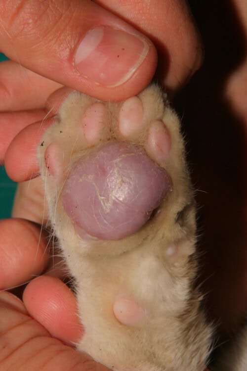
Almost exclusively affecting feline paw pads, plasmacytic pododermatitis manifests as characteristic spongy swelling of multiple paw pads, hence its name “pillow paw.” Pads develop a white, scaly, silvery appearance with distinctive crisscrossing streaks. Central metacarpal and metatarsal pads are regularly and more severely affected, although all pads can be involved.
Progression can include ulceration, secondary bacterial infections, pain, and lameness. Nodular or ulcerated lesions show a hemorrhagic tendency. Extra-digital manifestations have been documented, particularly in the form of concomitant plasmacytic stomatitis in two out of twenty-six patients in a retrospective study. Three cases presented nasal swelling, two being diagnosed as “ectopic plasmacytic pododermatitis” based on histopathological evaluation and therapeutic response.
Feline plasmacytic pododermatitis
Pathogenesis and Predisposing Factors
The pathogenesis remains partially elucidated, but constant hypergammaglobulinemia, marked tissue plasmacytosis, and response to immunomodulatory treatments strongly suggest an immune system dysfunction. The hypothesis of a cutaneous reaction pattern to multiple triggers, including infections, remains controversial.
A high incidence of feline immunodeficiency virus and feline leukemia virus infections has been reported in affected cats. One study showed positive immunohistochemistry for FeLV in paw pad biopsies from a cat seropositive for both retroviruses, suggesting a potential role of polyclonal B lymphocyte stimulation and impaired plasma cell function. An allergic etiology has also been proposed, with some cases showing seasonal recurrence with active lesions during warm seasons and spontaneous winter regression.
Proliferative and Necrotizing Otitis Externa
Pathophysiology and Clinical Presentation
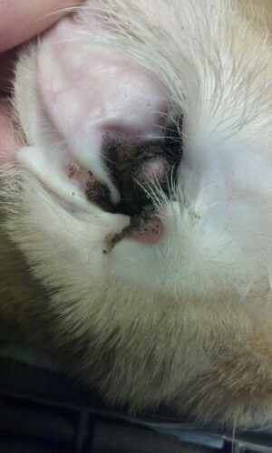
This rare condition is characterized by well-demarcated dark brown to black proliferative plaques covering the concave pinnae and extending into the vertical ear canal. Pathogenesis involves epidermal infiltration by CD3+ T lymphocytes inducing apoptosis of caspase-3 positive keratinocytes, although the origin of this lymphocyte activation remains unknown.
The plaques have a grainy, sand-like appearance and a friable consistency that favors bleeding upon manipulation. Accumulation of this friable material and thick, foul-smelling exudate can completely obstruct the ear canals. Secondary ear infections are a frequent complication. Extra-aural lesions, affecting the face with severe ulcerative and crusting dermatitis, tissue edema, and alopecia, have been described. Palpebral involvement manifests as dark keratinous debris with multifocal ulcers.
Proliferative and necrotizing otitis
Diagnostic and Therapeutic Modalities
Diagnosis relies on clinical features and histopathological examination. Biopsy of erythematous plaques should preserve adherent keratinous crusts to optimize diagnostic interpretation. Histology reveals severe acanthosis of the outer root sheath of hair follicles with scattered single-cell necrosis of keratinocytes at different epithelial levels.
Topical treatment with tacrolimus 0.1% twice daily is the initial reference therapy. Resolution can take three to twelve weeks, with no recurrence reported on two-year follow-up after treatment discontinuation. Topical corticosteroid monotherapy shows partial to no efficacy, although a combination of topical and oral corticosteroids has been reported as effective in an isolated case.
Oclacitinib, used off-label, has recently demonstrated remarkable efficacy in two cases treated at 1.5 mg/kg and 0.5 mg/kg orally twice daily, respectively. Complete remission was achieved in seven to twelve weeks. However, the safe therapeutic index in felines is not established, requiring close hematological monitoring given significantly lower levels of JAK2 in feline cells compared to canine cells.
Immune-mediated Alopecia
Pseudopelade
This rare immune-mediated condition is characterized by tropism of cytotoxic CD8+ T lymphocytes to the follicular isthmus. High titers of specific IgG autoantibodies directed against lower follicular structures, particularly trichohyalin and hair keratin, have been detected. Destruction of bulge stem cells leads to permanent alopecia.
Clinical presentation begins in adulthood with non-inflammatory and non-pruritic cicatricial alopecia, progressing over several months in a partially symmetrical bilateral pattern affecting the limbs, paws, abdomen, ventrolateral trunk, and face. The absence of broken hair shafts in affected areas distinguishes this condition from self-induced alopecia. Onychonyxis and onychomadesis may accompany the alopecia, indicating ungual matrix involvement.
Alopecia Areata
A dermatosis similar to alopecia areata has been described in a ten-year-old domestic short-haired cat presenting non-inflammatory alopecia of the ventral region and limbs, accompanied by onychomadesis. Histological and immunohistochemical examination revealed moderate to severe mural folliculitis and perifolliculitis at the follicular isthmus, composed of cytotoxic T lymphocytes. This localization differs from classic alopecia areata, defined by immune-mediated destruction of the follicular bulb rather than the isthmus.
Auricular Chondritis
Pathogenesis and Manifestations
This rare condition is characterized by inflammation and destruction of auricular cartilage, resulting from an immune-mediated process primarily targeting type II collagen. Pinnal lesions include swelling, thickening, deformation, pain, and intense erythema. Chronic progression can lead to purplish discoloration and curling of the pinnae. Bilateral involvement is the usual presentation.
Histopathological examination reveals inflammation, degeneration, necrosis, and loss of basophilic staining of the cartilaginous matrix, associated with perichondrial edema and fibrocyte and capillary endothelial proliferation. The cartilaginous inflammatory infiltrate is predominantly lymphocytic with some multinucleated giant cells.
Diagnosis of feline relapsing polychondritis requires histological documentation of chondritis in at least two different anatomical sites and/or involvement of at least two other organs. A case of a three-year-old Japanese domestic cat presented with auricular chondritis with costal, laryngeal, tracheal, and articular chondral modifications met these criteria, although the erosive polyarthritis suggests feline chronic progressive polyarthritis.
Therapeutic response is variable. Prednisolone at immunosuppressive doses can induce remission in three weeks, while dapsone at 1 mg/kg daily, with or without corticosteroids, has shown efficacy in four cases without recurrence after therapeutic discontinuation. Pinnectomy is a curative option in some situations. Spontaneous improvement and remission without treatment have also been reported.
Integrated Diagnostic and Therapeutic Approaches
Establishing the diagnosis of these immune-mediated dermatoses relies on combining clinical features, anamnesis, and histopathological examination. Cytology and skin biopsies are the most valuable diagnostic tools to differentiate these conditions from mimicking infectious, allergic, or neoplastic causes.
The existence of potential histological overlap between erythema multiforme and the SJS/TEN spectrum necessitates an integrated diagnostic approach. Microscopic pathological interpretation should be limited to a generic diagnosis of EM-TEN necrotizing epidermal disease, with subsequent subclassification depending on anamnesis, clinical signs, and lesion extent.
Therapeutic management varies depending on severity and presumed etiology. Immediate cessation of any suspicious drug is the absolute priority in SJS/TEN cases. Immunomodulatory treatments, including cyclosporine, mycophenolate mofetil, and corticosteroids, represent the therapeutic mainstays for most of these conditions. Clinical course and prognosis differ considerably depending on the pathological entity, with some conditions showing a tendency for spontaneous resolution while others require prolonged immunosuppressive treatment or constitute true dermatological emergencies.
Banovic F, Gomes P, Trainor K. Feline immune-mediated skin disorders: part 2. J Feline Med Surg. 2025;27:1-15.

