Immune-mediated skin diseases in cats, although rare, present a diagnostic and therapeutic challenge for veterinary practitioners. These pathologies, characterized by a dysregulation of the normal immune response, can manifest with variable clinical signs, ranging from erythema and alopecia to skin erosions, with or without pruritus, often mimicking other more common dermatological conditions.
Familiarity with the distinctive clinical characteristics of immune-mediated dermatoses is essential for enabling an early and accurate diagnosis, leading to appropriate therapeutic management. This article examines the main feline immune-mediated skin diseases, including the pemphigus complex, cutaneous lupus erythematosus, and exfoliative dermatoses associated or not with thymoma, detailing their clinical manifestations, diagnostic approaches, therapeutic options, and prognoses.
Feline Pemphigus Foliaceus
Pathogenesis and Presentation
Pemphigus foliaceus (PF), considered the most common autoimmune dermatosis in cats, is characterized by the binding of pathogenic autoantibodies to epidermal desmosomal adhesion proteins. This binding causes acantholysis and the recruitment of inflammatory cells, leading to the formation of vesicles in the superficial epidermis. Unlike humans and dogs, where the desmosomal targets of autoantibodies are well-characterized, investigations in cats are limited to the detection of anti-keratinocyte IgG by direct and indirect immunofluorescence. The desmosomal target remains currently unknown, but it is likely different from that in dogs due to the detection of positive immunostaining on oral mucosal tissue.
PF has been reported in different feline breeds (Siamese, Persian and Persian cross, Burmese, etc.) without specific racial or sexual predisposition. According to published case series, PF generally affects adult cats with a median age of 5-6 years, although the range varies between 5 months and 17 years depending on the studies.
Similar to canine PF, the majority of cats do not present with underlying triggering factors associated with the development of PF. Historically, a few rare cases of drug-induced PF have been reported, and a unique case simultaneously presented with exfoliative dermatitis associated with thymoma and PF as part of a paraneoplastic syndrome.
Diagnostic Approach
The diagnosis of feline PF currently relies on a combination of criteria including: (i) history and characteristic distribution of skin lesions, (ii) exclusion of other acantholytic neutrophilic pustular dermatoses (superficial staphylococcal pyoderma, pustular dermatophytosis), and (iii) cytology and/or histopathology confirming acantholytic pustular dermatitis.
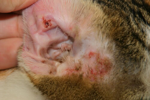
Erythematous macules and pustules represent the primary skin lesions of feline PF. However, due to their superficial epidermal localization, pustules are transient and rapidly evolve into erosions and crusts, often the most frequently observed lesions during physical examination. The most commonly affected areas include the head/face (nasal planum, eyelids, chin), ear pinnae, ungual skin folds, paw pads, and periareolar areas. When PF affects the ungual skin folds, significant crust formation is generally observed, with accumulation of purulent to caseous exudate and erosions that can progress to ulcerations.
Pruritus is reported in most cats with PF, and some patients present with systemic signs including lethargy, fever, weight loss, lymphadenopathy, and anorexia.
The presence of acantholysis in suspected PF lesions can be evaluated by cytology (standard Diff-Quik staining) from intact pustules, under recent moist crusts, or from purulent exudate in nail folds and/or by biopsy of these lesions. Classic cytology of PF lesions reveals the presence of acantholytic keratinocytes with a variable number of well-preserved neutrophils and/or eosinophils. Histopathological examination shows subcorneal or intragranular pustules with acantholytic keratinocytes and a perivascular to interstitial neutrophilic or mixed neutrophilic and eosinophilic infiltrate.
Clinically, pemphigus foliaceus can mimic allergic dermatitis
Treatment and Prognosis
Although the majority of cats with PF do not have an underlying trigger factor (e.g., medications), elimination of any suspected causal factor should be implemented immediately. Exposure to ultraviolet radiation has been associated with exacerbation of PF skin lesions in humans and dogs. Although no cases of UV-induced flare-ups have been reported in cats with PF, owners should be informed of this potentially aggravating factor.
The therapeutic management of feline PF remains challenging and generally requires immunosuppressive drugs to achieve clinical remission and long-term control of the disease. Oral glucocorticoid monotherapy has been considered the cornerstone of treatment for feline PF (prednisolone 2-4 mg/kg/day; triamcinolone acetonide 0.2-2 mg/kg/day; dexamethasone 0.1-0.2 mg/kg/day). Prednisone, a prodrug metabolized to active prednisolone, is not recommended in cats due to lower absorption and/or reduced conversion of prednisone to prednisolone.
Although most cats with PF achieve complete remission (absence of new lesions with healing of original lesions) within a few weeks of glucocorticoid monotherapy, only a minority of cats (4-15%) maintain this remission if glucocorticoid administration is discontinued. Therefore, corticosteroid-sparing adjuvants, such as cyclosporine (5-10 mg/kg/day) and chlorambucil (0.1-0.3 mg/kg/day), have been suggested to induce earlier clinical remission and ensure long-term control of PF.
In general, feline PF has a good prognosis, with most cats achieving complete remission with medical management (glucocorticoid monotherapy) within a median of 22-36 days. However, relapses under maintenance therapy are common, especially when treatment is reduced or discontinued. Unlike dogs, cats with PF are rarely euthanized due to progression of skin lesions despite treatment, adverse effects associated with treatment, or poor quality of life.
Pemphigus Vulgaris
Unlike PF, little information is available regarding feline pemphigus vulgaris (PV). The clinical and histopathological features of feline PV resemble those of canine and human PV; a similar pathological mechanism of desmosomal antibody targeting is therefore proposed for feline PV. Investigations in humans and dogs have identified desmoglein-3 as the main autoantigen. Currently, the targets of autoantibodies in feline PV skin lesions remain unknown.
Based on the few cases reported in the literature, flaccid vesicles are rarely observed in feline PV. In contrast, superficial erosions and ulcerations of mucocutaneous junctions constitute the main clinical characteristics. In reported cases, skin lesions frequently affect the lips, gums, hard palate, nasal planum, and philtrum; hairy skin lesions and/or paw pads are occasionally involved. Given the location of the lesions, lethargy, anorexia, halitosis, hypersalivation, and submandibular lymphadenopathy are commonly observed.
Definitive diagnosis relies on history, clinical signs, and skin biopsy, which shows suprabasal acantholysis, cleavage formation, and a “tombstone” arrangement of the basal layer. Multiple biopsies are necessary to capture diagnostic areas of PV, with intact vesicles and/or margins of erosions to ulcers with adjacent “normal” skin generally sampled.
Treatment of feline PV patients with oral glucocorticoids (4-6 mg/kg/day prednisolone) resembles the approach used in humans and dogs, and shows some success in controlling the disease. However, in refractory cases of feline PV, corticosteroid-sparing immunomodulators (chlorambucil, cyclosporine) should be considered, similarly to the treatment of feline PF.
Lupus Erythematosus
Cutaneous lupus erythematosus (CLE) may affect only the skin or present as part of a diverse array of life-threatening clinical signs in patients with systemic lupus erythematosus (SLE). Unlike humans and dogs, SLE and CLE variants like discoid lupus erythematosus (DLE) have rarely been published in cats.
A single case of SLE (anemia, thrombocytopenia, positive antinuclear antibodies) with signs of CLE presented with well-demarcated symmetrical alopecia, erosions to ulcerations, and crusts on the face, ears, neck, abdomen, limbs, and paw pads. Original reports on feline DLE described clinical signs of erythema, scaling, alopecia, erosions to ulcerations, and crusts with or without dyspigmentation (hyperpigmentation or depigmentation) affecting the head, ear pinnae, trunk, and paw pads; no more recent description has been published. In 2005, two adult cats with CLE presented with exfoliative dermatitis (alopecia, scaling) and erosions to ulcerations resembling the skin lesions observed in feline thymoma-associated exfoliative dermatitis.
In described feline SLE/CLE cases, histological examination of skin biopsies revealed a lymphocyte-rich interface dermatitis specific to CLE with vacuolar (hydropic) degeneration of basal keratinocytes, as well as lymphocytic mural interface folliculitis. Treatment of feline SLE/CLE patients resembles the approach used in humans and dogs, involving sun avoidance, use of topical glucocorticoids/tacrolimus for localized lesions, and systemic immunomodulatory drugs for generalized lesions.
Exfoliative Dermatitis Associated or Not with Thymoma
Thymoma-Associated Exfoliative Dermatitis
Thymoma-associated exfoliative dermatitis is a rare paraneoplastic syndrome where skin signs are frequently noted first, despite the probable initial presence of the neoplastic process. Non-cancerous skin lesions related to neoplasia occur at a site distinct from the primary tumor or its metastases. In cats, thymoma is the most common thymic neoplasia, originating from thymic epithelial cells in the cranial mediastinum.
The pathogenesis of feline thymoma-associated exfoliative dermatitis has not been elucidated, but an immune-mediated process similar to graft-versus-host disease is suspected. It has been proposed that thymoma-associated exfoliative dermatitis in cats results from a CD3+ T-cell mediated process caused by abnormal antigen presentation by neoplastic thymic epithelial cells that cross-react with epidermal keratinocytes.
This disease generally affects middle-aged to older cats, although it has been reported in cats as young as 4 years. No sex or breed predisposition has been identified.
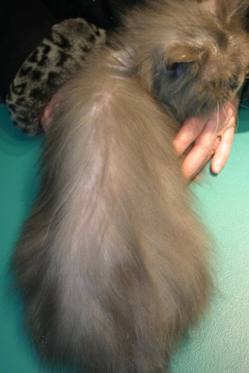
In cats with thymoma-associated exfoliative dermatitis, skin lesions first appear on the head and ear pinnae, then gradually progress to the back and trunk before becoming generalized. These areas gradually become scaly. Alopecia develops as exfoliation intensifies and lesions generalize in an asymmetric pattern. Brown, waxy, seborrheic debris accumulates in the nail folds and between the toes. Pruritus is not common, but affected cats may become mildly pruritic.
In addition to skin signs, cats with thymoma-associated exfoliative dermatitis may present with mild to severe lethargy and respiratory and gastrointestinal signs, such as dyspnea, cough, vomiting, and regurgitation. At presentation, these signs are generally proportional to the size of the mediastinal mass and intensify as the mass increases.
The diagnosis of thymoma-associated exfoliative dermatitis relies on history, clinical and histopathological findings, and the presence of a cranial mediastinal mass on imaging examination. Histopathology shows: marked orthokeratosis to focal parakeratosis; mild to moderate epidermal hyperplasia, with hydropic degeneration of basal keratinocytes and transepidermal apoptotic keratinocytes; a cell-poor to cell-rich interface dermatitis composed primarily of CD3+ lymphocytes with fewer plasma cells and a low number of mast cells and neutrophils.
Surgical excision of the tumor is the treatment of choice for the majority of cats suspected of thymoma-associated exfoliative dermatitis. The prognosis for animals with non-invasive and resectable thymomas is good, with skin lesions gradually resolving after tumor excision. In cats treated with surgical excision alone, an overall 3-year survival rate of 74% has been observed. In contrast, cats with invasive thymomas have higher recurrence rates and, postoperatively, mortality ranging from 11% to 22%.
Diffuse dorsolumbar alopecia related to a thymoma
Exfoliative Dermatitis Not Associated with Thymoma
A syndrome of exfoliative dermatitis similar to thymoma-associated exfoliative dermatitis has been reported in cats, but without a determined etiology. This condition is called exfoliative dermatitis not associated with thymoma because the skin lesions and histopathological examination are indistinguishable from cases of thymoma-associated exfoliative dermatitis. In cats that benefited from long-term follow-up, no development of thymoma was observed.
As with thymoma-associated exfoliative dermatitis, an immune-mediated process is suggested for exfoliative dermatitis not associated with thymoma, with CD3+ T-lymphocyte infiltration and epidermal cytotoxicity on histological examination.
For all cats with exfoliative dermatitis, imaging examination is crucial to confirm or rule out the presence of a cranial mediastinal mass, as the treatment protocol changes radically for exfoliative dermatitis not associated with thymoma.
Cats with exfoliative dermatitis not associated with thymoma respond to immunosuppressive treatment, and most patients require long-term treatment to maintain remission. The majority of cases achieve remission with modified cyclosporine (6.75-7.5 mg/kg every 24 hours) alone or in combination with prednisolone (2-4 mg/kg every 24 hours). Relapses may occur, particularly if immunosuppressive therapy is discontinued.
In conclusion, although immune-mediated skin diseases in cats are rare, they can be associated with severe systemic clinical signs, leading to poor quality of life and sometimes euthanasia. In-depth knowledge of the distinctive clinical characteristics of different immune-mediated skin disorders is essential to enable early and accurate diagnosis, as well as appropriate treatment.
Banovic F, Gomes P, Trainor K. Feline immune-mediated skin disorders – Part 1. J Feline Med Surg. 2025;27:1-13.

