Even though hyperadrenocorticism or Cushing’s syndrome is an endocrinopathy essentially described in dogs, it should not be forgotten that it can also be occasionally observed in cats or ferrets. However, in the latter species, it does not result from hypercortisolism, and therefore, it should not be referred to as Cushing’s syndrome.
Author: William Bordeau
Cabinet VetDerm,
1 avenue Foch 94700 MAISONS-ALFORT
In cats, nearly 80% of hyperadrenocorticism cases are of pituitary origin. However, spontaneous hyperadrenocorticism is very rare in cats. What is most commonly observed is iatrogenic Cushing’s syndrome, which results from the administration of corticosteroids or progestogens, although it is true that cats are more resistant than dogs in this regard.
Spontaneous Cushing’s syndrome appears in elderly to very elderly cats. The clinical signs that may be present are quite similar to those observed in dogs. Polydipsia, polyphagia, and weight loss may occur. In some cases, apathy and anorexia may be observed. The animal may have a pendulous abdomen, just as in dogs. Concomitant diabetes mellitus is present in nearly 90% of spontaneous hyperadrenocorticism cases, which necessitates biochemical and hematological analysis when Cushing’s syndrome is suspected. Chronic hypercortisolemia indeed exerts a diabetogenic action.
Furthermore, this diabetes mellitus is almost impossible to control without controlling the hyperadrenocorticism. Skin lesions are present in nearly 50% of cases. Alopecia, seborrhea, chronic or recurrent pyoderma, comedones, and hyperpigmentation may occur. Alopecia generally affects the abdomen, flanks, and thorax. In nearly 50% of cases, the skin is abnormally thin and tears easily. It is possible that cats with an adrenal tumor more frequently present skin fragility than those with a pituitary tumor. In cases of iatrogenic Cushing’s syndrome, the clinical signs are quite similar.
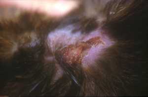
Skin fragility in a cat with iatrogenic Cushing’s
and flea allergy dermatitis
Regarding polyuria and polydipsia, the differential diagnosis notably includes renal failure or diabetes mellitus. The latter may appear independently or not of Cushing’s syndrome. Regarding dermatological manifestations, the differential diagnosis includes demodicosis, telogen effluvium, psychogenic dermatosis, hyperthyroidism, cutaneous asthenia, hepatic disease, or even a pancreatic tumor.
Anatomopathological analysis of skin biopsies is indicative of hyperadrenocorticism but does not allow for a definitive diagnosis. The latter can only be made through a complete endocrine exploration, which will begin, as we have previously seen, with a complete biochemical and hematological analysis. Although non-specific, leukocytosis, neutrophilia, lymphopenia, eosinopenia, hyperglycemia, hypercholesterolemia, and increased ALT activity may be observed. Unlike what is observed in dogs, alkaline phosphatase activity is only rarely increased. Subsequently, just as in dogs, an ACTH stimulation test or a dexamethasone suppression test can be performed.
The slight nuance in cats is that the dose of dexamethasone used in the low-dose suppression test corresponds to the dose used in the high-dose suppression test in dogs, which is related to the fact that cats are relatively refractory to administered corticosteroid doses. Just as in dogs, the measurement of the urinary cortisol to urinary creatinine ratio is somewhat uninteresting in cats. The search for a tumor or adrenal hyperplasia can be performed by ultrasound. Unfortunately, feline adrenal glands are difficult to observe, and this examination can therefore only be performed by an expert person and with good equipment.
Hyperadrenocorticism due to an adrenal tumor is the main cause of bilateral and symmetrical alopecia in ferrets. In nearly 84% of cases, it is unilateral. The left adrenal gland is more frequently affected. Unlike other species, polyuria, polydipsia, and polyphagia are rarely observed in this species. Similarly, few biochemical or urinary changes are generally noted. Basal cortisol concentration is usually within the normal range. The clinical manifestations of hyperadrenocorticism in ferrets are more similar to those observed in dogs with sex hormone-related dermatoses than those observed with cortisol overproduction, and for good reason.
Indeed, in cases of hyperadrenocorticism in ferrets, there is no overproduction of cortisol, but rather of sex hormones, particularly progesterone, androstenedione, and estradiol. Nearly 96% of ferrets with hyperadrenocorticism have at least one of these hormones elevated. In nearly 22% of cases, an elevation of all these hormones is even observed. Hyperadrenocorticism generally appears between 2 and 8 years of age and is manifested by bilateral, symmetrical, and non-pruritic alopecia that begins at the tail and progresses to the abdomen, inner thighs, and dorsolumbar region. In very advanced cases, this alopecia affects the dorsal neck region and the top of the head. The face and paws are generally spared.
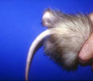
Alopecia localized to the tail and the base of the dorsolumbar region
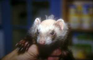
Alopecia on the top of the head, but the face remains spared
It is observed that 9 to 30% of animals exhibit pruritus. This does not respond to antihistamines and corticosteroids, and it is mainly localized between the shoulders. Comedones may be observed on the tail. Apart from these dermatological manifestations, apathy, splenomegaly, muscle atrophy, and vulvar swelling may be observed. Cases of mammary hyperplasia have been described. In nearly 19% of affected males with hyperadrenocorticism, difficulty urinating is noted. The enlarged adrenal gland can be palpated in almost 30% of cases.
The confirmatory diagnosis is made by ultrasonography. However, just as in cats, this examination must be performed by an expert person and with good equipment, as ferret adrenal glands are not easy to visualize. Thus, it is estimated that the adrenal tumor can only be observed in 50% of cases. Nearly 27% of affected ferrets also present with an insulinoma, hence the need to also observe the pancreas.
Endocrine exploration is difficult to perform in ferrets, and few laboratories perform the appropriate assays with established normal values in this species. It also appears that this exploration is rather disappointing in this species.
Finally, let us remember that even if these are relatively rare dysendocrinic dermatoses in our country, particularly concerning ferret hyperadrenocorticism, and unlike in the United States where it is much more frequent, these should not be neglected in these 2 species.
Bibliography
FeldmanE, Nelson: Canine and feline endocrinology and reproduction. 2nd edition. WB Saunders Co. Philadelphia, 1996.
Bruyette-DS. An approach to diagnosing and treating feline hyperadrenocorticism. Veterinary-Medicine. 2000, 95: 2, 142-148.
Zerbe-CA. Differentiating tests to evaluate hyperadrenocorticism in dogs and cats. Compendium, 2000, 22: 2, 149-158
Kemppainen EJ & coll. Endocrine responses of normal cats to TSH and synthetic ACTH administration. JAAHA, 1984, Vol. 20, pp 737-.
Lusson-D; Billiemaz-B Un cas d’hypercorticisme spontané chez un chat. Point Vét., 2000, 31: 204, 57-60
Rosenthal-KL; Peterson-ME; Bonagura-JD. Hyperadrenocorticism in the ferret. Kirk’s-current-veterinary-therapy-XIII:-small-animal-practice. 2000, 372-374.
Rosenthal-KL. Adrenal gland disease in ferrets.Vet.-Clin. N. Am.,-Small-Anim. Pract.. 1997, 27: 2, 401-418.
Gould WJ & coll. Evaluation of urinary cortisol:creatinine ratios for the diagnosis of hyperadrenocorticism associated with adrenal gland tumors in ferrets. JAVMA, 1995, Vol 206, pp 42-.
Rosenthal KL & coll. Hyperadrenocorticism associated with adrenocortical tumor or nodular hyperplasia of the adrenal gland in ferrets: 50 cases (1987-1991). JAVMA, 1993, Vol. 203, 271-.
A Muller, JF Quinton, V Chetboul, T Reviron, C Herbert, C Gau. Un cas d’hypercortisme chez un furet. Prat. Méd. Chir. Anim. Comp., 2001, Vol 36, Iss 1, pp 43-53


