During the last annual GEDAC conferences, our colleagues Anne Roussel and Pauline De Fornel had the opportunity to provide a comprehensive overview of oncology, from actinic keratosis to squamous cell carcinoma. This included a review of the latest pathogenic and therapeutic advancements.
The transition from actinic keratosis (AK) to feline cutaneous squamous cell carcinoma (SCC) represents a major area of interest in veterinary oncology, highlighting the importance of a thorough understanding of pathogenic mechanisms, accurate diagnosis, and appropriate therapeutic management. This common skin cancer in cats is intrinsically linked to ultraviolet (UV) light exposure and particularly affects light-coated animals. These conferences explored various facets of this pathology, from its precancerous origins to the most advanced therapeutic options.
Feline Cutaneous Squamous Cell Carcinoma, a Major Challenge in Veterinary Oncology
Cutaneous squamous cell carcinoma (SCC) is a widespread and potentially dangerous cancer in cats, primarily affecting the skin and mucous membranes. It is the third most common skin tumor in this species, representing approximately 10% of all feline cutaneous tumors, and up to two-thirds of eyelid tumors. The etiology of this neoplasia is strongly associated with environmental factors, particularly excessive exposure to ultraviolet (UV) rays, coupled with individual predispositions such as light coat color and advanced age. White-coated cats, for example, have a more than 13 times higher risk of developing cutaneous SCC compared to colored cats. Although feline cutaneous SCC is characterized by significant local aggressiveness, often invasive or proliferative, its metastatic potential is generally limited, estimated at approximately 5% of cases, with the majority of metastases observed in regional lymph nodes. This behavioral profile of the tumor underscores the imperative of early diagnostic and therapeutic management to optimize the prognosis and maintain an adequate quality of life for the animal.
Actinic Keratosis: The Precancerous Prelude to Cutaneous Squamous Cell Carcinoma
Actinic keratosis (AK), also known as solar keratosis, is a precancerous skin lesion that frequently precedes the onset of cutaneous squamous cell carcinoma. Its development is the result of the cumulative effect of UV rays on the skin, highlighting the importance of chronic sun exposure throughout the animal’s life.
Definition and Etiology of Actinic Keratosis
In human medicine, AK is recognized as a major public health problem in sunny regions, such as Australia, where its prevalence is notable. Predisposing factors include skin phototype (Fitzpatrick classification), age (adults over 60 years in human medicine, cats over 9 years for SCC), and sex (men being more frequently affected, possibly due to less sun protection). In our feline companions, as well as in dogs and horses, AK also results from excessive sun exposure. White cats are particularly predisposed to AK and SCC, as are sparsely haired areas of the body.
Pathogenesis and Molecular Mechanisms of Transformation
The underlying mechanism of actinic keratosis formation and its progression to squamous cell carcinoma is complex and involves UV-induced genetic alterations. UV rays are classified into three categories: UVC,
normally filtered by the ozone layer; UVB, known to be the most
erythemogenic spectrum; and UVA, which have a deeper action and are considered the most damaging, particularly in the 320 to 340 nanometer wavelength. This cumulative UV exposure leads to direct and indirect damage to the genetic material of keratinocytes. UVAs produce reactive oxygen species, such as superoxide anions, which cause oxidative damage to nucleic acids. UVBs, on the other hand, induce nucleotide base mismatches in DNA, leading to the formation of mutagenic photoproducts such as pyrimidine dimers, and thus mutations. A key player in protection against this damage is the P53 gene, a tumor suppressor gene. When genetic material is altered, P53 can either induce apoptosis (programmed cell death) if the damage is irreparable, or halt the cell cycle to allow DNA repair. However, mutations in the P53 gene are frequently observed in AK and SCC lesions, and even in perilesional areas, which compromises this protective function and promotes the clonal expansion of precancerous and cancerous keratinocytes. UV exposure also promotes the production of arachidonic acid, a pro-inflammatory mediator, which is converted into prostaglandins by cyclooxygenases (COX). Prostaglandins are potent mediators of inflammation that promote cell proliferation, tumor invasion, angiogenesis (formation of new blood vessels supplying the tumor), and the growth of tumor cells, and even metastases. Overexpression of cyclooxygenase 2 (COX-2) has been demonstrated in AK and SCC lesions in humans, laboratory animals, cats, and dogs. Finally, epigenetics, meaning the influence of lifestyle and environment (such as sun exposure) on gene expression, plays a crucial role. Genes involved in precancerous and cancerous lesions can be modified throughout an individual’s life, influencing the maturation of lesions and their progression to cancer. Actinic keratosis can evolve in three ways: spontaneous regression (in 25% of cases in human medicine), stability, or unfortunately progression to invasive squamous cell carcinoma. The early stages of actinic keratosis are considered reversible if sun exposure is effectively limited. Beyond this stage, the lesions are no longer reversible, hence the critical importance of early detection for prevention.
Clinical Manifestations and Diagnosis of Actinic Keratosis
Actinic keratosis lesions are preferentially located on sparsely haired and sun-exposed areas, such as the nasal planum, ear pinnae, temporal regions, eyelids, lips, and abdomen, particularly in white and blue-eyed cats who are highly predisposed. Initially, AK may present as a solar dermatitis, comparable to a degenerating sunburn, with erythema and some scales, accompanied by hair loss. These lesions tend to worsen from summer to summer, making the skin increasingly vulnerable. Gradually, the lesions may become more crusty, more depilated, erythematous, sometimes with curling of the free edge of the ear pinna or nasal planum. Cutaneous horns, indicating a proliferation of the superficial layers of the epidermis, may also be observed. Ulcerated lesions may appear, often due to pruritus and scratching, which makes clinical distinction from early squamous cell carcinoma difficult. The differential diagnosis of actinic keratosis includes various skin conditions such as dermatophytosis, sunburn, balanitis, hypersensitivity reactions, autoimmune phenomena such as pemphigus, and Notoedric mange. Due to the complexity of clinical diagnosis and the need to distinguish these lesions from an infiltrative neoplastic process, performing cytology and, imperatively, a biopsy is crucial to confirm the diagnosis and guide management. Histopathological examination allows observation of epidermal dysplasia, actinic dermatoses, hyperplasia, and alteration of dermal elastic fibers. The basement membrane remains preserved until the stage of carcinoma in situ, but is destroyed in infiltrative forms, thus marking progression to SCC.
Feline Cutaneous Squamous Cell Carcinoma: An Aggressive Oncological Entity
Squamous cell carcinoma (SCC) is a malignant tumor derived from keratinocytes, the cells that make up the superficial layer of the skin. It is characterized by pronounced local aggressiveness, but relatively low metastatic potential, although regional lymph node metastases are possible.
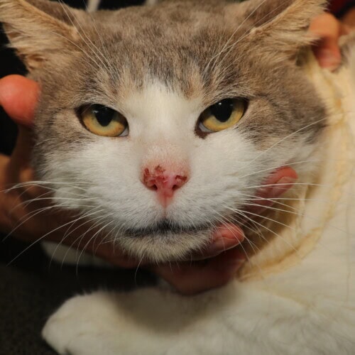
Squamous cell carcinoma of the nasal planum
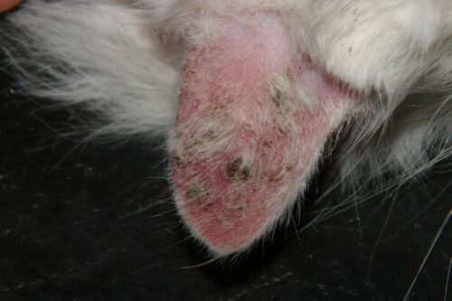
Diffuse ear involvement in a white cat
Epidemiology and Risk Factors for Feline Cutaneous Squamous Cell Carcinoma
As previously mentioned, SCC is a common cancer in cats, representing the third most common skin tumor. A predisposition in light-coated cats, and particularly white cats, is a well-recognized risk factor due to their increased sensitivity to ultraviolet (UV) rays. Chronic sun exposure is the primary etiological factor, with UV rays causing cumulative genetic damage. Advanced age (generally over 9 years) is also an important risk factor, as are genetic predispositions and, in some cases, the involvement of certain viruses such as papillomavirus. Lesions most often appear on hairless or poorly pigmented areas of the face, including the nasal planum, bridge of the nose, eye canthi, ear pinnae, and lips. Multiple facial lesions are frequently observed, in approximately 30% of cases.
Clinical Manifestations and Tumor Classification
The clinical signs of SCC are varied and can evolve with the progression of the disease. Initially, lesions may present as a crust with peripheral erythema, frequently progressing to an ulcerated, invasive lesion. Other alarming signs include persistent ulcers that do not heal, localized hair loss, persistent scabs, chronic wounds, and changes in skin color. These tumors are locally very aggressive, often infiltrative and destructive of surrounding tissues. Although the metastatic potential is limited (approximately 5%), regional lymph node metastases are the most common. A study on feline oral squamous cell carcinomas even revealed lymph node metastases in 31% of cats and pulmonary metastases in 10% of cases. The World Health Organization (WHO) tumor classification is essential for prognostic evaluation and therapeutic decision-making. It distinguishes stages based on tumor infiltration and size:
-
Tis : in situ
-
T1 : superficial, < 2 cm
-
T2 : slightly infiltrative, 2-5 cm
-
T3 : subcutaneous infiltration, > 5 cm, or infiltration of fascia, muscles, cartilage, bone
-
T4 : any size with deeper infiltration. Tumor size is a fundamental prognostic factor, with small lesions associated with much more favorable therapeutic outcomes. For example, for a truffectomy, the median survival is 16 months for tumors of stage < T2, compared to only 5 months for tumors > T3.
Specific Forms of Feline Squamous Cell Carcinoma
SCC can develop in various locations, each with specific clinical and prognostic characteristics:
Ear Squamous Cell Carcinoma
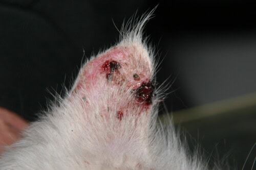
With 72% of cases, the ear is the most frequently affected area, particularly the edges or base of the ear. Symptoms include swelling or ulceration, crusts or bleeding, itching, scratching, head shaking, pain, and weight loss. Although local infiltration is often limited, the prognosis is poor if the cancer is already advanced.
Oral Squamous Cell Carcinoma
This is the most frequent oral tumor in cats. This type is particularly aggressive and can spread rapidly to neighboring structures. The most frequent locations are the maxilla (38% of cases), tongue/pharynx, mandible, lips, cheek, or tonsil. The prognosis varies according to the location: better for cheek or tonsil lesions (median survival of 724 days), but poorer for caudal pharyngeal involvement (survival less than 50 days). Symptoms include pain or difficulty eating, loss of appetite and weight, excessive salivation, bleeding from the mouth, facial or jaw swelling, and the presence of a visible mass or ulcer. The extension workup for oral forms is crucial and includes fine-needle aspiration of mandibular lymph nodes (even of normal size) and thoracic radiographs. CT scans are also essential to evaluate underlying osteolysis, which is very common in this pathology.
Diagnosis of Feline Cutaneous Squamous Cell Carcinoma
The definitive diagnosis of SCC relies on a methodical approach. A thorough clinical examination is the first step, allowing the identification of suspected masses or skin anomalies. However, confirmation of the malignant nature of the tumor imperatively requires a biopsy of the lesion, followed by histopathological examination under the microscope. To assess the extent of the disease and detect possible metastases, complementary imaging techniques are used:
-
Radiographs and ultrasounds can be used to assess local and regional spread.
-
CT scans (computed tomography) are particularly useful, even essential, for large or infiltrative tumors, especially to assess the extent of underlying osteolysis in oral forms.
-
Fine-needle aspirates of regional lymph nodes (especially mandibular) are recommended in cases of suspected lymphadenomegaly (enlargement of lymph nodes).
Therapeutic Strategies: A Personalized Approach for Cats with SCC
The therapeutic management of feline cutaneous squamous cell carcinoma is complex and must be individualized, considering the local aggressiveness of the tumor and the need to preserve the animal’s quality of life. The key to favorable outcomes lies in early management of the lesion. The choice of therapeutic modality depends on several objective and subjective criteria, including the location and volume of the tumor, the cat’s general condition, the extent of the disease (although its importance is less for cutaneous SCC), the availability and cost of treatments, as well as aesthetic considerations and owner motivation. A diagnostic confirmation by biopsy is essential before initiating any radical treatment.
Surgery: The Curative Treatment of Choice
Radical surgical excision of the lesion is often the first-line treatment when technically feasible and accepted by the owner. The primary goal of surgery in oncology is to achieve a cure by complete removal of the tumor with clear margins.
Case of ear lesions: Pinna amputation is considered a relatively simple surgical procedure and is most often curative, especially if the intervention occurs before the entire pinna is infiltrated. Although this may cause aesthetic damage, it is generally better accepted by owners for ear lesions than for other locations.
Case of nasal planum lesions: Truffectomy, i.e., amputation of part of the nasal planum, is also a preferred option for small lesions (Tis and T1 stages). It allows radical surgery with a curative objective. Median survival after truffectomy is approximately 22 months. The aesthetic results, although modified, are often deemed acceptable by owners, especially after being reassured by comparative photos.
Limitations of surgery: Radical surgery is not always possible or appropriate. Locations such as the temples or eyelids pose significant challenges due to the complexity of necessary reconstructions and the difficulty of ensuring clear margins. For eyelid lesions, early radical surgery might involve associated enucleation to ensure no recurrence. Similarly, a significant tumor volume can make radical surgery impossible, even for usually favorable locations. If the surgical excision margins are infiltrated, there is a risk of recurrence, and adjuvant treatment is then recommended. In situations where radical surgery is not feasible, cytoreductive surgery can be performed, but it must be supplemented with adjuvant treatment such as radiotherapy.
Radiotherapy: An Effective Local Control Option
Radiotherapy is a treatment of choice for squamous cell carcinoma, particularly when surgical excision is incomplete, for inoperable tumors, or when surgery is refused by the owner. It aims to destroy tumor cells by irradiation. Several radiotherapy techniques are available:
Contact and Interstitial Radiotherapy (Brachytherapy)
Strontium-90: Used for very superficial tumors (less than 2 mm deep), especially those of Tis and T1 stages. This technique delivers a localized radiation dose and offers very good results, with remission in 90% of cats at one year and 80% at two years in some studies. Side effects are rare. However, its availability in France is limited.
Curietherapy (Iridium-192, Cobalt)
Involves applying a radioactive source directly to the tumor. Historically, “iridium wires” were used, allowing dosimetry adapted to the tumor volume with excellent results. Due to radiation protection constraints, this technique has evolved towards high-dose-rate brachytherapy, where the radioactive source is handled remotely. The results are very encouraging, with more than 96% responses (including 72% complete responses) and a median remission duration of approximately 10 months, as well as satisfactory aesthetic results. Marseille is one of the few centers in France to use a cobalt source for this approach.
External Beam Radiotherapy (Electrons and Photons)
Administered from outside the body by particle accelerators. For superficial tumors, electrons are preferred due to their limited penetration, allowing maximum dose deposition in the first millimeters or centimeters of tissue.
Hypofractionated protocols: Involve a reduced number of sessions (e.g., 4 to 5 fractions) with high doses per session, spaced approximately one week apart. The results are honorable, with about 50% responses and a median remission of 9 months.
Accelerated hypofractionated protocols: Represent the most recommended approach currently for feline SCC. They consist of numerous sessions administered over a very short period (e.g., twice a day, Monday to Friday, over one week). These protocols offer very satisfactory results, with 94 to 100% complete responses and long remission durations (13 to 30 months, or even several years). Acute side effects are generally well tolerated.
Stereotaxy
A more recent technique using one to two sessions with very large doses. Preliminary results are encouraging, but acute side effects are notable.
Orthovoltage
Another form of external beam radiotherapy using lower energy radiation (kilovolts), suitable for superficial lesions. It is available in Brive la Gaillarde and the Paris region.
The main centers offering external beam radiotherapy in France include Lille, the Nantes school, and Créteil. Important prognostic factors in radiotherapy are tumor size (small tumors achieving better results) and Ki67 (a cell proliferation marker). The side effects of radiotherapy almost systematically include hair loss and depigmentation of the treated area, and are more significant for large volume tumors.
Electrochemotherapy (ECT): Potentiation of Chemotherapy Effects
Electrochemotherapy (ECT) is an innovative technique that aims to increase the chemosensitivity of tumor cells, even those considered poorly chemosensitive. It combines the administration of a cytotoxic agent with the local application of electrical impulses.
Principle: After intravenous (or sometimes intratumoral) injection of a cytotoxic agent (historically bleomycin, but carboplatin is used in France because bleomycin is not authorized for this indication), an electric current is applied directly to the tumor a few minutes later. These electrical impulses temporarily increase the membrane permeability of tumor cells, allowing better incorporation of the active principle and, consequently, potentiation of its local effects.
Protocols and Results: The number of sessions generally varies between one and four, often one or two are sufficient. The oncology team at VetAgro Sup reports very promising results with carboplatin: 80% complete responses after a single session and 100% after a possible second session, with only one recurrence observed after 11 months in 9 cats. Larger studies report between 65% and 96% complete responses, with a median remission of between 4 and 36 months. ECT is considered a safe, well-tolerated, and very effective technique.
Prognostic Factors and Side Effects: As with radiotherapy, tumor volume is a major prognostic element: early-stage tumors (Tis, T1) show more favorable results in terms of remission duration, and local side effects (ulceration, bleeding) are significantly less for small tumors.
Availability and Cost: ECT is available in a greater number of centers in France than certain radiotherapy techniques, which can influence the choice. Its cost is undoubtedly lower than that of radiotherapy.
Photodynamic Therapy: A Targeted Approach for Early Lesions
Photodynamic therapy is a technique that relies on the topical application of a photosensitizer to the tumor, followed by illumination with an intense red light.
Indications and Results: This method is primarily indicated for in situ forms and small tumors (Tis to T2 stages). It allows for high complete response rates, ranging from 85% to 100% after a single treatment. However, remission durations are generally shorter (median of 3 to 5 months), with 20% to 60% recurrence. The possibility of performing new sessions in case of recurrence is an advantage. Aesthetic results are considered perfect.
Side Effects and Availability: Local side effects are rare and limited. This technique is less documented in veterinary medicine than other options, and its availability in France is restricted, being more common in Germany or Italy.
IV.E. Other Treatments and Supportive Care
Systemic Chemotherapy
Systemic chemotherapy alone has not shown significant efficacy for the treatment of feline cutaneous SCC, as these tumors are considered poorly chemosensitive.
Cryotherapy and Laser Therapy
These procedures can be used to destroy tumor tissue in some cases and are less invasive than traditional surgery. They are generally reserved for infiltrative, nodular, or evolving lesions.
Systemic Retinoids
The efficacy of systemic retinoids is still to be confirmed and remains less documented in veterinary medicine.
Non-Steroidal Anti-Inflammatory Drugs (NSAIDs)
Topical diclofenac 3% (Solaraze®) can be used locally to manage pruritus and pain. Although studies in dogs have shown that firocoxib (a selective COX-2 inhibitor) can normalize epidermal keratinocyte proliferation, evidence of the efficacy of coxibs in the context of feline SCC is not conclusive, and their use should be done with great caution due to possible side effects (e.g., kidney failure).
Topical Imiquimod
This immunomodulator is used for the treatment of actinic keratoses and early squamous cell carcinomas. It is applied two to three times a week for six weeks. Although it can cause an intense local inflammatory reaction, with initially painful crusts and ulcerations, clinical results are often spectacular, with complete disappearance of lesions in a few weeks. The owner must wear gloves during application.
Supportive and Palliative Care
Regardless of the chosen treatment, supportive care is essential to improve the cat’s quality of life throughout its illness. This includes rigorous pain management, control of secondary infections, and appropriate nutritional support. When healing is no longer an option, palliative care aims to relieve symptoms and maintain the animal’s comfort.
Prevention and Early Detection: The Pillars of Effective Management
Prevention and early detection are the most effective strategies to combat feline cutaneous squamous cell carcinoma.
Prevention of Sun Exposure
Given the predominant role of UV rays in the etiology of SCC, limiting sun exposure is paramount, especially for light-coated cats.
Sun Avoidance: It is recommended to avoid outings and direct sun exposure during peak UV intensity hours, generally between 10 AM and 5 PM. It is also crucial to limit prolonged naps behind windows, as even glass does not completely block UVA. The installation of filtering films on windows can be considered.
Specific Sunscreens for Animals: The application of sunscreens adapted for animals is an important complementary measure. Two types of filters are distinguished:
-
Physical filters: Composed of titanium dioxide and zinc oxide, they form an opaque barrier that reflects UV rays. They are water-resistant but can make the coat greasy and be potentially toxic if ingested in large quantities. They require fewer reapplications than chemical filters.
-
Chemical filters: More transparent, they are stored in the stratum corneum and absorb UV rays. However, they decompose under the effect of solar radiation and require more frequent applications (at least 3 to 4 times a day).
In general, physical filters are preferred in animals due to a limited number of applications and the risk of licking.
Physical Protections: The use of protective clothing can be an option in some cases.
Owner Education: It is essential to inform owners of white cats, from a young age and during vaccination consultations, about the risks associated with sun exposure and the preventive measures to adopt.
Early Detection
Owner vigilance and regular veterinary examinations are essential for detecting precursors or early lesions of SCC.
Monitoring for Signs: Owners should be alert to the appearance of swelling, changes in skin or mucous membrane color, unexplained loss of appetite or weight, or behavioral changes. Any ulceration that does not heal, persistent crust, or chronic wound should prompt a prompt veterinary consultation.
Veterinary Examinations: Regular visits to the veterinarian allow for early screening of any suspicious lesions. Rapid diagnosis significantly expands available treatment options and improves the chances of therapeutic success and the cat’s quality of life.
Conclusion
Feline cutaneous squamous cell carcinoma is a common and often locally aggressive neoplasm, whose genesis is closely linked to cumulative sun exposure and precancerous actinic keratosis lesions. Understanding the pathogenic mechanisms, risk factors, and clinical manifestations is fundamental for a rigorous diagnostic and therapeutic approach. The key to effective treatment lies in early detection and management of the disease. Radical surgery remains the treatment of choice for small and well-localized tumors, offering the possibility of cure. However, when surgery is not feasible or is refused by the owner, other therapeutic modalities have demonstrated their effectiveness. Radiotherapy, in its various forms (brachytherapy, external beam radiotherapy, particularly accelerated protocols), and electrochemotherapy, especially with carboplatin, represent effective options for local tumor control, offering high response rates and significant remission durations. Photodynamic therapy also offers remarkable aesthetic results for very early lesions. The choice between these different techniques must be adapted to each individual case, considering the location and volume of the tumor, the cat’s general condition, comorbidities, the availability of treatments, and the owner’s preferences. In addition to curative or palliative treatments, supportive care is essential to improve the patient’s quality of life. Finally, primary prevention through limiting sun exposure and applying adequate protection, as well as secondary detection through regular veterinary examinations and careful monitoring of skin lesions, remain the most effective strategies to reduce the incidence and morbidity associated with this oncological pathology in cats.


