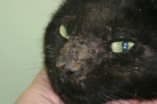
Pemphigus Foliaceus (PF) stands out as the most frequent form of pemphigus observed in domestic cats. It is also the most commonly diagnosed autoimmune dermatosis in this species. This skin condition, while often presenting highly suggestive clinical and cytological manifestations, nevertheless requires histopathological confirmation to establish a definitive diagnosis.
Over the years, the therapeutic management of feline PF has undergone significant evolution, mainly due to a better understanding of its pathogenesis and the emergence of new drug options. These advances have considerably improved the prognosis and quality of life for cats affected by this condition.
Pathogenesis
Pemphigus represents a complex set of diseases that affect a variety of species, including not only humans and cats, but also dogs, horses, and goats. In all these species, the fundamental pathogenic mechanism relies on an autoimmune attack targeting the intercellular connections between keratinocytes in the granular layer of the epidermis. More specifically, specific immunoglobulins bind to these cell junctions, causing their destruction and leading to the separation of keratinocytes. This process, known as acantholysis, leads to the detachment of keratinocytes from the underlying epidermal layers.
Once released, these keratinocytes undergo a characteristic morphological transformation: they round up and take on a peculiar appearance. These cells, now called “acantholytic keratinocytes” or “acantholytic cells,” are distinguished by their intensely stained cytoplasm and intact nucleus. It is interesting to note that, unlike in humans and dogs where specific desmosomal targets have been identified, the exact target of this autoimmune attack in cats remains to be elucidated. This particularity highlights the complexity of feline PF pathogenesis and emphasizes the need for further research in this area.
The pathological process does not stop there. In response to the attack on adhesion molecules, inflammatory cells, mainly neutrophils, invade the epidermis at the lesioned sites. This cellular infiltration leads to the formation of pustules, which are the characteristic primary lesions of PF. These pustules, although transient, play a crucial role in the clinical and cytological diagnosis of the disease.
Epidemiology
The epidemiology of feline PF presents interesting characteristics. Unlike some other feline dermatological conditions, PF does not appear to show a clear predisposition related to the age, breed, or sex of affected cats. This absence of an obvious predisposing factor makes the disease potentially relevant for all cats, regardless of their individual characteristics.
To illustrate this point, it is useful to refer to the two largest retrospective studies conducted to date on feline PF. The first, which examined 57 cases of cats with PF, revealed a wide range of disease onset ages, ranging from less than one year to 17 years, with a median age of 5 years. These results were corroborated by a more recent study of 49 cats, which reported similar onset ages, ranging from 5 months to 15 years, with a slightly higher median age of 6 years.
This extensive age distribution highlights the importance for veterinarians to consider PF as a potential differential diagnosis in cats of all ages presenting compatible clinical signs. Furthermore, the absence of racial or sexual predisposition means that all cats, regardless of their breed or sex, should be considered susceptible to developing this disease.
Etiology
The etiology of PF in cats remains largely enigmatic. In the majority of cases, the underlying cause could not be determined with certainty. However, clinical observations and case studies have raised the possibility that some cases of feline PF may be drug-induced. This hypothesis has opened up a new field of investigation in understanding the disease.
The mechanism by which drugs could induce PF is complex. It is believed that certain drugs could directly trigger the activation of proteolytic enzymes in the skin. These enzymes, once activated, would attack desmosomes, the structures responsible for adhesion between keratinocytes. This attack would lead to acantholysis, the characteristic pathological process of PF.
Among the suspected drugs, methimazole has received particular attention. Several cases of feline PF have been attributed to the use of this drug, commonly prescribed to treat hyperthyroidism in cats. It is particularly interesting to note that the histopathological appearance of methimazole-induced lesions is indistinguishable from that observed in non-drug-induced PF cases. This similarity highlights the complexity of diagnosis and the need for a thorough drug history in cats suspected of PF.
Other drugs have also been implicated in possible induced PF cases. These include cimetidine, an H2 receptor antagonist used to treat stomach ulcers, ampicillin, a broad-spectrum antibiotic, itraconazole, an antifungal, and ipodate, a radiological contrast agent. Although these associations have been reported, it is important to note that a causal link has not been definitively established in all cases.
The possibility of drug-induced PF underscores the importance of a detailed history when evaluating a cat presenting clinical signs consistent with PF. Veterinarians should be particularly attentive to any recent changes in the cat’s medication, including the introduction of new drugs or changes in dosage.
Clinical Characteristics
Primary and Secondary Lesions
The clinical picture of feline PF is characterized by a progressive evolution of skin lesions. The earliest lesion of feline PF may be an erythematous macule, although this initial phase is rarely observed in clinical practice. Indeed, the progression of the disease is often rapid, and owners usually only notice symptoms when the disease has already reached a more advanced stage.
The pustular phase, which rapidly follows the macular phase, is more characteristic and more easily identifiable. The pustules of feline PF have distinctive characteristics that differentiate them from those observed in other skin conditions, such as bacterial folliculitis. Unlike bacterial pustules, which are generally centered on a single hair follicle, PF pustules extend over several hair follicles. This difference is crucial for differential diagnosis.
However, it is important to note that these pustules are extremely fragile and transient. Their ephemeral nature means that they rupture quickly, evolving into crust formation. These crusts, typically honey-colored, are dry and can be irregular and confluent. They often constitute the most obvious lesion during clinical examination.
Facial crusty lesions
Beneath these crusts, the condition of the skin can vary. In some cases, intact, though alopecic and scaly, skin may be found. However, it is more common to observe erosions under the crusts. These erosions result from the rupture of pustules and the loss of the superficial layers of the epidermis.
Pruritus may sometimes be present
Lesion Distribution
The distribution of lesions in feline PF follows a relatively predictable pattern, although individual variations may occur. The two largest retrospective studies conducted to date have identified the most frequently affected areas.
The ear pinna is often the first and most consistently involved area. Lesions on the ears can be particularly striking and are often what first catches the attention of owners.
The rest of the head and face is also frequently affected. This includes the periorbital area, the nose and muzzle, and the chin. These locations can significantly impact the cat’s appearance and well-being, as they can interfere with important functions such as vision and eating.
The paws are another commonly affected area. Lesions can develop on all parts of the paws, including the paw pads and the interdigital region. In some cases, paw involvement can be severe enough to cause lameness.
The trunk, both dorsal and ventral, is generally less frequently involved than the previously mentioned areas. However, it is important to note that study results differ slightly on this point. One study reported that the trunk was affected approximately half as often as the paws, while another found more frequent trunk involvement. This variability highlights the importance of a thorough clinical examination, as lesions can appear in unexpected areas.
A particular point to note is periareolar involvement, observed in approximately a quarter of cases. This localization can be easily overlooked if the examination is not meticulous, hence the importance of a complete inspection of the cat’s entire body surface.
A crucial aspect of lesion distribution in feline PF is their bilaterally symmetric nature. This symmetry is a distinctive characteristic of the disease and can help differentiate it from other skin conditions. All studies agree on this point, emphasizing its diagnostic importance.
Systemic Signs
Although PF is primarily a skin disease, it may be accompanied by systemic signs in some patients. These signs can vary in intensity and frequency, but their presence can significantly impact the cat’s general well-being and disease management.
Pruritus is one of the most commonly observed signs. Its intensity can range from mild to moderate. In the two largest retrospective studies, pruritus was present in 66% to 80% of cats with PF. This itching can not only be uncomfortable for the cat, but can also lead to secondary lesions due to excessive scratching and licking.
Lethargy is another frequently reported sign. Affected cats may appear less active, less interested in their surroundings, or spend more time sleeping than usual. This fatigue may be due to the discomfort caused by skin lesions or be a sign of systemic inflammation.
Fever is also observed in some patients. It can be intermittent or persistent and often reflects the general inflammatory state associated with the disease. The presence of fever may require additional management and may influence treatment selection.
Less frequently, some cats may present with anorexia and weight loss. These signs can be particularly concerning as they can lead to a rapid deterioration of the cat’s general condition. Anorexia may be due to general discomfort, pain associated with oral lesions if present, or a side effect of medications used to treat the disease.
Lymphadenopathy has also been reported in some cases. Enlargement of lymph nodes can be localized, corresponding to the most affected skin areas, or generalized, reflecting a systemic immune response.
In cases where the claws and paw pads are severely affected, lameness may be observed. This lameness can vary in intensity and may affect one or more paws. It can be due to pain associated with the lesions or inflammation of surrounding tissues.
It is important to note that the presence and intensity of these systemic signs can vary considerably from patient to patient. Some cats may only present with skin signs, while others may show a combination of skin and systemic signs. This variability underscores the importance of a comprehensive clinical evaluation and regular follow-up of patients with PF.
Bibliography
Anderson, JG; Bizikova, P; Linder, KE | Erosive and ulcerative stomatitis in dogs and cats: which immune-mediated diseases to consider? | J Am Vet Med Assoc. 2023 Apr 17;261(S1):S48-S57. doi: 10.2460/javma.22.12.0573. PMID: 37059419
Barrs, VR; Beatty, JA; Hobi, S; Sandy, JR | Successful management of feline pemphigus foliaceus with pentoxifylline and topical hydrocortisone aceponate. | Vet Med Sci. 2022 May;8(3):937-944. doi: 10.1002/vms3.768. PMID: 35212177
Bizikova, P; Burrows, A | Feline pemphigus foliaceus: original case series and a comprehensive literature review. | BMC Vet Res. 2019 Jan 9;15(1):22. doi: 10.1186/s12917-018-1739-y. PMID: 30626385
Bizikova, P; Levy, BJ; Mamo, LB | Detection of circulating anti-keratinocyte autoantibodies in feline pemphigus foliaceus. | Vet Dermatol. 2020 Oct;31(5):378-e100. doi: 10.1111/vde.12861. PMID: 32372490
Bizikova, P; Mendoza-Kuznetsova, E; Piedra-Mora, C | Comorbidity of ectopic thymoma-associated exfoliative dermatitis and pemphigus foliaceus in a cat. | Can Vet J. 2021 Oct;62(10):1067-1070. PMID: 34602633
Izydorczyk, V; Pye, C | Pemphigus foliaceus in cats. | Can Vet J. 2024 Mar;65(3):297-300. PMID: 38434171
Jordan, TJM; Affolter, VK; Outerbridge, CA; Goodale, EC; White, SD | Clinicopathological findings and clinical outcomes in 49 cases of feline pemphigus foliaceus examined in Northern California, USA (1987-2017). | Vet Dermatol. 2019 Jun;30(3):209-e65. doi: 10.1111/vde.12731. PMID: 30779233
Linder, KE; Olivry, T; Tham, HL | Deep pemphigus (pemphigus vulgaris, pemphigus vegetans and paraneoplastic pemphigus) in dogs, cats and horses: a comprehensive review. | BMC Vet Res. 2020 Nov 23;16(1):457. doi: 10.1186/s12917-020-02677-w. PMID: 33228633
Related searches
feline pemphigus foliaceus, autoimmune disease, foliaceus in cats, pemphigus foliaceus in, autoantibodies, immune system, female cat, muzzle, animals, dermatology, dermatophytosis, dermatoses, onychitis, vesicles, pyoderma, demodicosis, consultation, relapses, dogs, chow chow, screws, dai, mucous membranes, constituents, cases of pemphigus foliaceus, mechanisms, side effects, article, most, course, in dogs, neutrophils, number, stratum corneum, biopsy, sensitivity, contact, photos, pinnae, group, proteins, components, neutrophilic granulocytes, feline pemphigus foliaceus, autoimmune disease, autoimmune, pemphigus, cat, dermatosis, foliaceus in cats, pemphigus foliaceus in, autoantibodies, pemphigus foliaceus, in cats, dog, pf, cases, management, scabs, skin, level, horse, muzzle, immune system, female cat, lesions, disease, well-being, ears, paws, owners, age, feline, chin, onychitis, form, problems, appearance, areas, dai, screws, side effects, constituents, animals, mechanisms, consultation, chow chow, keratinocytes, diagnosis, treatment, presence, species, clinical signs, neutrophils, contact, blisters, anamnesis, components, group, family, desmogleins, membrane, clinical examination, proteins, condition, muzzle, quality, options, cells, diseases, symptoms, evolution, history, itching, pustules

