At the last American Congress of Veterinary Dermatology held in Orlando, Florida, our colleague Paul Bloom had the opportunity to provide a comprehensive update on non-pruritic alopecias in dogs, presenting his complete diagnostic approach.
July 2025
Indeed, the systematic approach to non-pruritic alopecias in dogs often presents a diagnostic challenge for the veterinary practitioner. By methodically addressing each case, it is first necessary to determine whether the condition is congenital or acquired, and then to evaluate its focal or diffuse distribution. These initial observations effectively narrow down the possibilities, thereby optimizing the clinical approach.
Causes of non-pruritic alopecia notably include ischemic dermatopathies, sebaceous adenitis, various endocrinopathies, and follicular dysplasias. For each of these entities, a rigorous diagnostic approach and adapted therapeutic strategies must be implemented, taking into account both clinical elements and owner expectations.
Initial Diagnostic Approach
The examination of a dog with non-pruritic alopecia requires a structured and methodical approach. When faced with an animal showing hair loss without associated itching, the first step is to determine whether the condition is congenital or acquired. This fundamental distinction immediately directs the diagnostic process towards specific pathological groups.
If the alopecia is acquired, the next step is to establish whether its distribution is focal/multifocal or symmetrical/diffuse. This topographic differentiation constitutes a major decision point in the diagnostic algorithm for canine alopecias. Indeed, the underlying causes differ considerably according to the observed distribution pattern.
Evaluation of Focal to Multifocal Alopecia
When faced with focal to multifocal alopecia, the first questions to ask are:
-
Are there papules, pustules, or epidermal collarettes?
-
If yes, and if these lesions are follicular, consider:
- Pyoderma
- Demodicosis
- Dermatophytosis
- And in the case of follicular pustules, also consider pemphigus
-
-
-
In the absence of such lesions, it is necessary to determine if the alopecia is self-induced.
- If so, think of allergies or parasites
- If not, consider a biopsy to look for inflammatory, structural, cyclical, or neoplastic problems.
-
This methodical approach significantly narrows the field of differential diagnoses and avoids unnecessary investigations. The presence of primary lesions like papules and pustules generally suggests an infectious or inflammatory process, while their absence tends to point towards structural or dysfunctional processes affecting the hair cycle.
Excluding Prior Causes
Before considering a biopsy for focal to multifocal alopecia, it is imperative to systematically exclude the following causes, which can be ruled out by a good history and/or immediate additional examinations:
- Congenital causes
- Pruritic causes
- Infectious causes
This rigorous diagnostic approach avoids premature invasive explorations and guides towards more specific and effective treatments. Complementary examinations such as skin scrapings, trichograms, and fungal cultures should be performed prior to any biopsy to exclude demodicosis and dermatophytosis, which are common causes of focal to multifocal alopecias.
Ischemic Dermatopathies
Ischemic dermatopathies constitute a significant group of causes of focal to multifocal alopecia in dogs. They are characterized by lesions resulting from altered cutaneous vascularization, thereby compromising blood supply to the hair follicles and other skin structures.
Definition and Classification
Two main categories are distinguished based on their histopathological presentations:
- Vasculitis: characterized by the presence of inflammatory cells in the vessel wall.
- Vasculopathy: marked by vascular damage without significant inflammation.
Histopathologically, vasculopathy is characterized by a loss of endothelial cells and thickening of vascular walls, without significant inflammatory cell infiltration. These lesions essentially represent the imprint of a resolved prior inflammatory process, leaving vascular sequelae. They are “vascular scars” indicative of a past inflammatory episode.
Dermatomyositis
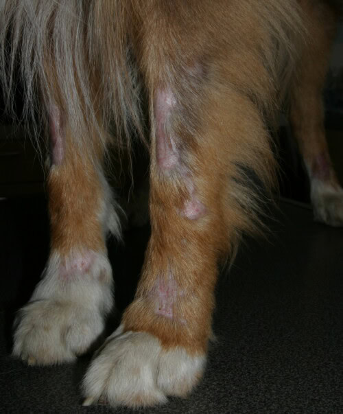
Dermatomyositis is a genodermatosis primarily affecting Collies and Shelties. Onset usually occurs between six weeks and one year of age, but signs are typically observed before six months of age. It is an autoimmune inflammatory disease affecting both the skin and muscles.
Dermatomyositis
Clinical Presentation
Skin lesions include:
- Focal to multifocal areas of alopecia
- Scales and crusts
- Potential erosions and ulcers
- Variable depigmentation and hyperpigmentation
- Possibly infarct-like necrosis, especially on the ears
- Scars
- Petechiae and purpura
Characteristic distribution includes the face, mucocutaneous junctions, tail and ear tips, and pressure points such as elbows and hocks. Carpal and tarsal involvement, truncal alopecia, and sometimes onychodystrophy can also be observed.
Muscular manifestations, although less common, can be proportional to the severity of skin lesions. They include atrophy of masticatory and locomotor muscles, and sometimes megaesophagus. This muscle involvement generally occurs after the development of skin lesions.
Vaccination-induced Alopecia
A particular case of ischemic dermatopathy is vaccination-induced alopecia, primarily associated with the rabies vaccine. Lesions generally appear 2 to 12 months after vaccine administration and more frequently affect small white breeds.
The implicated mechanism is a type III hypersensitivity reaction, where a soluble antigen binds to IgG or IgM, forming immune complexes that deposit in small vessels. These immune complexes cause local or distant vascular inflammation.
This phenomenon explains why lesions can appear distant from the injection site – typically at the ear tips despite injection elsewhere on the body. The route of administration (subcutaneous or intramuscular) does not influence the occurrence of this reaction.
Clinical lesions consist of focal (sometimes multifocal) areas of alopecia, scales, plaques, hyperpigmentation, nodules, erosions, crusts, and skin atrophy (scars). Histopathologically, in addition to typical vasculitis changes, septal panniculitis and focal lymphoid nodules with pale blue material in the subcutaneous tissue are observed, likely representing persistent vaccine adjuvant.
Idiopathic Vasculitis
Idiopathic vasculitis can occur in any breed and may present as a localized or generalized form. The distinction between a generalized form associated with a vaccine and a generalized idiopathic form primarily relies on anamnesis, particularly the history of recent vaccination.
The histopathological peculiarities of the vaccine-associated form include:
- Septal panniculitis
- Focal lymphoid nodules
- Pale blue material suggestive of persistent adjuvant in the subcutaneous tissue
Potential Triggers
Before classifying vasculitis as idiopathic, it is essential to rule out all potential triggers, including:
- Infectious causes (bacteria, demodicosis, tick-borne diseases, heartworm disease, viral infections)
- Other immune-mediated diseases like discoid lupus erythematosus
- Food allergies
- Medications and vaccines
- Neoplasias, which constantly release antigens that can form immune complexes
Any foreign antigen to which the dog is exposed can potentially trigger antibody formation and, if it is a soluble antigen forming complexes with IgG or IgM, lead to vasculitis by immune complex deposition in small vessels. The presence of eosinophils on biopsy may suggest food allergy as a potential trigger.
Diagnosis
The diagnosis of ischemic dermatopathies relies on:
- Signalment
- Physical examination
- Characteristic histopathological changes
Histopathology is generally essential for a definitive diagnosis. It is important to note that focal lesions in young dogs can resemble other conditions such as demodicosis, focal pyoderma, dermatophytosis, or discoid lupus. In the case of dermatomyositis, breeders often mistakenly attribute facial lesions to trauma from playing with littermates or a household cat.
Therapeutic Approach
Regarding the monitoring of subclinical urinary tract infections in dogs on corticosteroids or immunosuppressants, recommendations have evolved. Contrary to previous beliefs, these dogs can develop a bladder inflammatory response despite immunosuppression, as evidenced by the ability to develop pyoderma (subcutaneous pus) while on steroids. Approximately 10-15% of healthy dogs and up to 30% of dogs with chronic diseases may have asymptomatic bacteriuria that does not require treatment. The International Society for Companion Animal Infectious Diseases has published specific recommendations on the appropriate management of urinary tract infections.
Sebaceous Adenitis
Sebaceous adenitis constitutes another major cause of non-pruritic alopecia in dogs, with distinct clinical and histopathological characteristics.
Pathogenesis and Epidemiology
Sebaceous adenitis is an inflammatory disease specifically targeting the sebaceous glands. It has a well-identified autosomal recessive genetic basis in the standard Poodle. However, it can affect many other breeds without its hereditary nature being formally demonstrated in these cases.
Commonly affected breeds include:
- Standard Poodle (confirmed autosomal recessive form)
- Akita
- Samoyed
- Bichon Havanese
- English Springer Spaniel
- Old English Sheepdog
- Belgian Shepherd
- Various other spaniel breeds
Prevalence seems to be increasing with better recognition of the disease by clinicians. Sebaceous adenitis typically affects young to middle-aged dogs.
Some authors distinguish two forms of the disease:
- The granulomatous form (Standard Poodle form) observed in Standard Poodles, Akitas, Samoyeds, and Shepherds.
- The short-haired breed form seen in Vizslas, Weimaraners, and Dachshunds – although this is considered by some to be more related to sterile granuloma/pyogranuloma syndrome (sterile peri-adnexal granulomatous dermatitis) rather than sebaceous adenitis itself.
Clinical Presentation
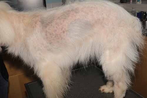
The characteristic form in the Standard Poodle manifests as:
- Adherent white scales
- Follicular casts (keratinous debris adhering to the hair shaft, visible emerging from the follicular ostium)
- Scaling within the hair follicle that fails to shed normally
- Varying degrees of hypotrichosis potentially leading to alopecia
- Dull coat
- In dogs where hair regrows, loss of the typical Poodle curls
These follicular casts resemble candle wax that has dripped onto a wick. When present, the differential diagnosis should include sebaceous adenitis itself, but also superficial bacterial folliculitis, demodicosis, and dermatophytosis.
Secondary infections are common due to follicular obstruction, and can induce pruritus through an inflammatory response.
An interesting recent discovery concerns the association of sebaceous adenitis with ophthalmic abnormalities. Affected dogs may develop ocular disease related to a thinner tear film, as Meibomian glands are actually a modified form of sebaceous glands. Manifestations include:
- Red eyes
- Ocular discharge
- Dry eyes
This complication is reported to occur in approximately 50% of affected dogs, potentially warranting a Schirmer tear test and ophthalmic examination, even in the absence of clinical signs reported by the owner.
Sebaceous adenitis in a Samoyed
Lesion Distribution
The typical distribution of sebaceous adenitis begins on the head and progresses caudally and distally. This topographical evolution is characteristic and can help guide diagnosis.
In the short-haired breed form (which may constitute a distinct entity), multifocal annular areas of alopecia with scales involving the trunk are observed.
Diagnosis
Diagnosis of sebaceous adenitis relies on:
- Characteristic clinical presentation
- Exclusion of other causes of alopecia
- Histopathological confirmation
Early histopathological changes in the granulomatous form include a nodular granulomatous to pyogranulomatous reaction in the ischemic region of the hair follicle, with unilateral localization (sebaceous glands being unilateral), follicular and superficial hyperkeratosis (clinically visible as scales). In the terminal stage of the disease, inflammation resolves, giving way to perifollicular fibrosis, follicular atrophy, and the absence of sebaceous glands.
Differential diagnoses to consider when follicular casts are observed include:
- Sebaceous adenitis itself
- Superficial bacterial folliculitis
- Demodicosis
- Dermatophytosis
Therapeutic Approach
Illustrative Clinical Case
The case of Baxter, a dog affected by both sebaceous adenitis and allergies, well illustrates the complexity of these situations. Initially presented for his allergies, the sebaceous adenitis was not specifically treated because:
- The owner was not concerned about the dog’s appearance
- The dog had no odor
- He had no secondary pyoderma
After desensitization for his allergies, spontaneous improvement of the sebaceous adenitis was observed, although no causal correlation can be established with certainty. This case highlights that temporal correlation does not necessarily equal cause-and-effect, and that the absence of specific treatment can sometimes be a reasonable option when the disease does not affect the animal’s quality of life.
Symmetrical/Diffuse Alopecias and Endocrinopathies
When alopecia presents with a symmetrical or diffuse distribution, the diagnostic approach differs significantly from that of focal/multifocal alopecias.
Initial Evaluation
When faced with symmetrical or diffuse alopecia, it is essential to evaluate:
- The presence of constitutional signs
- The existence of hematological changes
Constitutional signs to look for include:
- Lethargy
- Heat seeking
- Polyuria/polydipsia
- Abdominal distension
- Hepatomegaly on palpation
- Bradycardia
Relevant hematological changes include:
- Mild non-regenerative anemia
- Hypercholesterolemia
- Elevated alkaline phosphatase
If constitutional signs are present, endocrinopathies should be actively investigated, including:
- Hypothyroidism
- Hyperadrenocorticism
- Hyperestrogenism (sometimes associated with a Sertoli cell tumor)
In the absence of systemic signs, a skin biopsy may be considered, although its diagnostic utility may be limited in some cases of symmetrical/diffuse alopecias.
Hypothyroidism
Hypothyroidism is often over-diagnosed in dogs. Many practitioners withdraw more dogs from thyroid supplementation than they put on treatment.
Clinical Presentation
Hypothyroidism most commonly results from immune-mediated destruction of the thyroid gland. It primarily affects medium to large breed dogs of middle age. Dermatological signs of hypothyroidism include:
- Alopecia
- Hyperpigmentation
- Triangular area of alopecia caudal to the nasal planum (also observed in spayed/neutered dogs)
- “Frizziness” in some breeds like Golden Retrievers and Irish Setters
- Dry or greasy seborrhea
- Poor hair regrowth (more common complaint than spontaneous alopecia)
- Recurrent bacterial pyoderma
- Dry, dull coat
Diagnostic Pitfalls
Several pitfalls should be avoided in the diagnosis of hypothyroidism:
- Do not test for hypothyroidism if the dog is pruritic (pruritus can cause post-inflammatory hyperpigmentation and self-induced alopecia)
- Interpret therapeutic trials with thyroid supplementation cautiously:
- Check blood levels after one month to confirm they are within the therapeutic range (upper end of reference interval or slightly above)
- Clinically evaluate the dog after three months to determine if therapy has had a positive impact
- Discontinue treatment if no clinical improvement is observed
Diagnostic Tests
Useful thyroid tests include:
- Total T4 (TT4)
- Free T4 by equilibrium dialysis (fT4ed)
- Canine TSH (cTSH)
- Anti-thyroglobulin autoantibodies (TgAA)
- Anti-T4 autoantibodies (T4ab)
- Anti-T3 autoantibodies (T3ab)
A complete thyroid profile should include TT4, cTSH, TgAA, T4ab, T3ab. fT4ed can be added in the presence of anti-T4 antibodies, non-thyroidal illness, or if the dog has received medications affecting thyroid function.
Important: dogs should not have received topical or oral corticosteroids for at least 30 days, or depot steroids for 3 months prior to testing. Sulfonamides should also be avoided for at least 30 days.
Influence of Non-Thyroidal Illnesses
Non-thyroidal illnesses can affect free and total T4 levels, complicating diagnosis. It is generally best not to test thyroid function in a sick dog, unless myxedema coma or another thyroid emergency is suspected.
The more severe the illness, the greater the divergence between total and free T4 may be, although this difference is not necessarily statistically significant. Rather than worrying about this phenomenon, it is simply better to wait until the dog is healthy to assess its thyroid function.
Treatment
For dogs diagnosed with hypothyroidism, the standard treatment consists of administering L-thyroxine at 0.02 mg/kg twice daily, preferably using brand-name medications over generics. After one month of treatment, a blood sample should be taken 4-6 hours after tablet administration to measure total T4. Levels should be in the upper part of the reference interval, or even slightly above.
Hyperadrenocorticism (Cushing’s Syndrome)
Hyperadrenocorticism is considered by some specialists to be a more frequently encountered endocrinopathy than hypothyroidism, both in its iatrogenic and spontaneous forms.
Atypical Presentation
Contrary to common belief, many dogs with hyperadrenocorticism do not present with classic signs such as:
- Polyuria/polydipsia
- Abdominal distension (“pot-belly”)
- Elevated liver enzymes
These “atypical” dogs may present solely with:
- Recurrent pyoderma
- Deep pyoderma
- Demodicosis
- Woolly or inappropriate coat
- Alopecia
- Calcinosis cutis (sometimes as the sole clinical sign)
- Comedones
This partial clinical presentation is frequently observed by dermatologists, but often unrecognized by general practitioners who look for the complete clinical picture of the disease. This “dermatological” form of hyperadrenocorticism poses a significant diagnostic challenge.
Diagnostic Approach
If hyperadrenocorticism is suspected:
- Proceed with a standard evaluation (CBC, biochemistry, urinalysis)
- Look for subtle indicators like a normal low urine specific gravity or a low normal BUN
- If the clinical appearance suggests hyperadrenocorticism despite the absence of biological abnormalities, continue investigations.
Important: first test for hyperadrenocorticism before hypothyroidism, as steroids decrease thyroid values (euthyroid sick syndrome).
Diagnostic Tests
Recommended tests include:
- ACTH stimulation test if recent steroid exposure
- Low-dose dexamethasone suppression test in the absence of recent steroid exposure
- Note: a normal test does not definitively rule out the disease
The author considers the sensitivity of the low-dose dexamethasone suppression test to be much higher than that of the ACTH stimulation test. The urine cortisol/creatinine ratio is considered less reliable, particularly in dogs presenting primarily with dermatological signs.
Treatment
The treatment of hyperadrenocorticism is determined by the severity of clinical signs. Therapeutic options include:
- Trilostane
- Mitotane
The choice between these molecules depends on several factors, including the type of hyperadrenocorticism (pituitary or adrenal), comorbidities, and the clinician’s experience with these medications.
Dyscyclic Follicular Diseases
Dyscyclic follicular diseases represent a group of conditions characterized by a structurally normal hair follicle but with an abnormality in the follicular cycle.
Classification and Etiology
These conditions receive different names depending on the affected breeds:
- Alopecia X
- Post-clipping alopecia
- Seasonal flank alopecia
Despite their varied names, these conditions share a common characteristic: hair does not grow normally due to a disruption of the hair cycle. The exact pathophysiological basis remains poorly understood.
Before making a diagnosis of dyscyclic follicular disease, it is essential to rule out known endocrinopathies (hypothyroidism, hyperadrenocorticism, hyperestrogenism) that may present similar clinical pictures.
Histopathological Evaluation
To differentiate these conditions, a skin biopsy can be performed with:
- An elliptical incision including the affected area and the adjacent clinically normal area
- A specific request for end-to-end sectioning to allow observation of disease progression
However, the diagnostic utility of biopsy is limited because the histopathological abnormalities of dyscyclic follicular diseases often resemble each other. Typical histological features include follicular atrophy, “telogenization” of follicles with excessive trichilemmal hyperkeratinization (flame follicles), orthokeratotic hyperkeratosis, follicular keratosis, and sebaceous gland atrophy.
These changes, although specific, do not allow differentiation between the various dyscyclic diseases, nor can they definitively distinguish them from endocrinopathies.
Alopecia X
Alopecia X primarily affects long-haired breeds and poodles. Its etiology remains mysterious, with several theories:
- Imbalance of adrenal sex hormones
- Abnormal hormonal metabolism at the follicular level
- Problem of hormonal receptors at the follicular level
The latter theory is supported by the observation that hair regrows at biopsy sites, suggesting a local rather than systemic inhibition of the hair cycle.
Clinical Presentation
Affected dogs gradually lose their guard hairs, usually starting from the neck and progressing to the shoulders, trunk, and thighs. The coat may become woolly, cream-colored, and in some cases, alopecia is accompanied by hyperpigmentation.
Diagnosis
Diagnosis relies on signalment, history, physical examination, and exclusion of other alopecic diseases. Histopathology can support the diagnosis but is not specific. A sex hormone adrenal stimulation test can be performed, but its diagnostic value is debatable.
Treatment
Seasonal Flank Alopecia
Seasonal flank alopecia primarily affects short-haired dogs like Boxers, Airedales, and Bulldogs.
Clinical Characteristics
This non-scarring alopecia has several peculiarities:
- Onset often in autumn with spontaneous resolution in spring (but sometimes the reverse)
- May occur only once or recur annually (sometimes with increasingly extensive areas)
- May persist without complete resolution
- Lesions typically on the flanks and sometimes on the caudolateral thorax
- Alopecia generally bilateral with annular lesions that can merge into polycyclic lesions
- Hyperpigmentation and smooth, shiny skin
- Possibility of developing papules and pustules consistent with secondary bacterial pyoderma
Etiological Theories
The exact etiology remains unknown. Some suggest a “melatonin deficiency” since many dogs develop lesions in the autumn, when melatonin levels should normally increase, and some respond to melatonin administration. However, this hypothesis cannot explain cases where hair is lost in spring and regrows in autumn.
Diagnosis and Classification
The classification of seasonal flank alopecia remains controversial:
- Some consider it a follicular dysplasia (structural abnormality of the hair follicle)
- Others interpret it as a hair cycle problem
- Histopathological examination reveals both abnormal hair follicles and cycle abnormalities
Diagnosis relies on excluding other non-scarring alopecias – a history alone can be diagnostic if it is a recurring problem. Biopsy can support but not definitively confirm the diagnosis.
Treatment
Treatment is based on:
- Natural evolution (frequent spontaneous resolution)
- Melatonin, more often used preventatively just before the onset of seasonal symptoms
Given that the disease generally goes into spontaneous remission, it can be difficult to determine if melatonin had a real impact, especially during the first occurrence.
Post-clipping Alopecia
Post-clipping alopecia primarily affects Nordic breeds.
Etiological Hypotheses
Two main theories explain this phenomenon:
- These breeds have a very long telogen (rest) phase to preserve their proteins; if the hair is cut during this phase, it will not regrow until the next anagen cycle.
- Clipping would decrease blood flow to the area (heat conservation mechanism), thereby reducing local growth factors.
Diagnosis and Treatment
Diagnosis relies on history and exclusion of endocrinopathies. Histopathology generally reveals normal-sized follicles but predominantly in the telogen phase.
Treatment may include:
- Patience (natural evolution)
- Sometimes short-term thyroid supplementation (7-10 days) to stimulate anagen formation
- A 90-day trial with melatonin
Structural Follicular Dysplasias
Structural follicular dysplasias are distinguished from dyscyclic diseases by the presence of anomalies not only of the hair follicle but also of the hair shaft.
Diagnostic Criteria
To diagnose a structural follicular dysplasia, histopathology must show both:
- Dysplastic hair follicles
- Dysplastic hair shafts
A 1998 study revealed that 46% of dogs with endocrine alopecia had dysplastic hair follicles, but less than 1% simultaneously had dysplastic hair shafts. This distinction is crucial for differentiating structural dysplasias from endocrine alopecias.
Color-Linked Dysplasias
This category includes:
Color Dilution Alopecia (CDA)
- Affects blue or fawn coated dogs (resulting from the “dilution” gene effect on black or brown hairs)
- Particularly affects Dobermans and Great Danes
- Autosomal recessive genodermatosis
- Dog born with normal coat then development of hypotrichosis/alopecia between 4 months and 3 years
- Only affects diluted color areas
- Dull coat, scales, and comedones
- Frequent secondary bacterial pyoderma
- Probable origin in dysfunctional melanin transfer from melanosomes to hair matrix or a defect in melanin storage
- Result: melanin aggregation weakening the hair shaft until fracture
Diagnosis relies on:
- History and physical examination
- Hair appearance on trichogram (melanin aggregation, architectural disruption)
- Exclusion of other causes of alopecia (demodicosis, dermatophytosis, pyoderma, endocrinopathies)
- Histopathological confirmation
Treatment primarily aims at:
- Removing the affected animal from breeding
- Managing secondary pyoderma and seborrhea
- Using baths, humectants, fatty acids ± antibiotics
- Potentially melatonin (6 mg three times daily for 90 days)
Black Hair Follicular Dysplasia (BHFD)
- Affects two-tone or three-tone coated dogs (Boston Terriers, Basset Hounds, Cocker Spaniels)
- Autosomal recessive transmission
- Similar mechanism to CDA (defective melanin transfer causing its aggregation)
- Anomalies usually noted at weaning, starting with a dull coat affecting only black hairs
- Progression to alopecia
- Possible secondary pyoderma
- Considered a localized form of CDA
- Treatment identical to CDA
Dysplasias Not Linked to Color
These dysplasias have been described in several breeds including:
- Portuguese Water Dogs
- Irish Water Spaniels
- Curly-Coated Retrievers
Between 6 months and 6 years of age (depending on breed), these dogs develop symmetrical hypotrichosis progressing to alopecia, generally starting on the neck and progressing to the shoulders, trunk, tail, and thighs. Remaining truncal hairs may change color (lightening).
An interesting pathophysiological element is the presence of estrogen receptors in telogen hair follicles, which are important for maintaining this phase. In Irish Water Spaniels, a dietary change (avoiding soy containing phytoestrogens) proved effective. Melatonin and trilostane, which block estrogen receptors, could explain their effectiveness in various canine alopecias.
Pattern Alopecia
Pattern alopecia is also a late-onset genodermatosis. Dogs are born with a normal coat but develop this alopecia between 6 months and 1 year of age.
There are four distinct forms of this non-inflammatory and non-pruritic alopecia:
-
Form affecting male Dachshunds:
- Slow progressive alopecia and hyperpigmentation of the ear pinnae
-
Form primarily observed in females of several breeds (Dachshunds, Chihuahuas, Whippets, Manchester Terriers, Greyhounds, Italian Greyhounds):
- Identical to the first form but with a different distribution
- Progressive alopecia caudal to and involving the ear pinnae
- Affecting the ventral neck, abdomen, and caudomedial thighs
-
Form observed in American Water Spaniels and Portuguese Water Dogs
- See description of non-color-linked dysplasias above
-
Form observed on the caudolateral thighs of Greyhounds
Regardless of the form, diagnosis relies on signalment, history, physical examination, exclusion of other alopecic diseases, and is confirmed by histopathology showing miniaturization of hair follicles and shafts with normal adnexa. Improvements have been reported in some dogs treated with melatonin.
General Therapeutic Approach to Non-Pruritic Alopecias
The therapeutic approach varies considerably depending on the specific diagnosis, but some common principles can be identified.
Basic Principles
- Identification of the underlying cause: Seeking the “due to” is essential to avoid ineffective symptomatic treatment.
- Consideration of owner expectations: Evaluating the importance placed on cosmetic appearance versus management of secondary complications.
- Individual adaptation: Personalizing treatment based on each patient and the owner’s ability to implement it.
Common Treatments for Multiple Conditions
Monitoring and Adjustment
Long-term management of non-pruritic alopecias requires:
- Regular evaluation of treatment efficacy
- Progressive and cautious adjustments of doses and frequencies
- Particular attention to potential side effects
- Constant communication with the owner
Differential Diagnosis and Systematic Approach
To effectively address non-pruritic alopecias in dogs, a systematic approach is essential.
Decision Algorithm
1/ Initial Evaluation:
Determine if the alopecia is congenital or acquired
If acquired, determine if it is focal/multifocal or symmetrical/diffuse
2/ For focal/multifocal alopecias:
Look for primary lesions (papules, pustules, epidermal collarettes)
Determine if the alopecia is self-induced
Exclude infectious and parasitic causes
3/ For symmetrical/diffuse alopecias:
Look for constitutional signs
Evaluate hematological changes
If present, orient towards endocrinopathies
If absent, consider dyscyclic or structural follicular diseases
Indications for Skin Biopsy
Skin biopsy is indicated in several situations:
- Focal/multifocal alopecia after exclusion of infectious and parasitic causes
- Alopecia not responding to standard treatments
- Suspicion of ischemic dermatopathy
- Doubt between several differential diagnoses
- Confirmation of a structural follicular dysplasia
For an optimal biopsy:
- Perform an elliptical incision including affected and normal areas
- Request a full end-to-end section
- Specify the search for specific signs based on clinical suspicions
Differential Diagnosis of Non-Pruritic Focal/Multifocal Alopecias
The main causes of non-pruritic focal to multifocal alopecias are:
1/ Ischemic dermatopathies
Dermatomyositis
Vaccination-induced alopecia
Idiopathic vasculitis
2/ Sebaceous adenitis
Standard Poodle form
Specific forms in other breeds
3/ Cutaneous neoplasms
Cutaneous lymphomas
Mast cell tumors
Other infiltrative skin tumors
4/ Localized non-inflammatory dermatophytosis
Sometimes a fungal infection can present as non-pruritic alopecia
Post-traumatic or post-inflammatory scars
Sequelae of previous inflammation or trauma
Differential Diagnosis of Non-Pruritic Symmetrical/Diffuse Alopecias
The main causes of non-pruritic symmetrical/diffuse alopecias are:
1/ Endocrinopathies
Hypothyroidism
Hyperadrenocorticism
Hyperestrogenism
2/ Dyscyclic follicular diseases
Alopecia X
Post-clipping alopecia
Seasonal flank alopecia
3/ Structural follicular dysplasias
Color-linked alopecias (CDA, BHFD)
Non-color-linked alopecias
Pattern alopecias
Atypical Manifestations of Non-Pruritic Alopecias
Certain atypical presentations deserve special attention:
1/ Mixed forms
Association of pruritic and non-pruritic alopecias
Secondary superinfections inducing pruritus in initially non-pruritic alopecia
2/ Dermatological hyperadrenocorticism
Form with predominant skin manifestations without classic systemic signs
Diagnosis often delayed by the absence of typical signs (PU/PD, distended abdomen)
3/ Partial dyscyclic follicular diseases
Affecting only certain body areas
May misleadingly suggest a focal distribution
Conclusion
The approach to non-pruritic alopecias in dogs requires a rigorous and systematic methodology. The distinction between focal/multifocal and symmetrical/diffuse distributions constitutes the first crucial step in the diagnostic algorithm. For each type of alopecia, specific causes must be considered and systematically ruled out.
Ischemic dermatopathies, sebaceous adenitis, endocrinopathies, and follicular dysplasias represent the main etiological categories of these conditions. Each has its own clinical, histopathological, and therapeutic characteristics.
Understanding the underlying mechanisms of these alopecias optimizes the diagnostic approach and adapts treatment to each patient. Identifying the “due to” – the fundamental cause – remains the primary objective, although this is not always possible, particularly in dyscyclic alopecias like alopecia X.
Communication with the owner is also essential, especially for conditions where the impact is primarily cosmetic. Treatment personalization must consider not only the specific diagnosis but also the owner’s capabilities and expectations.
Advances in understanding the pathophysiological mechanisms of these conditions pave the way for more targeted therapeutic approaches, although many questions remain unanswered regarding the precise etiology of certain non-pruritic alopecias.

