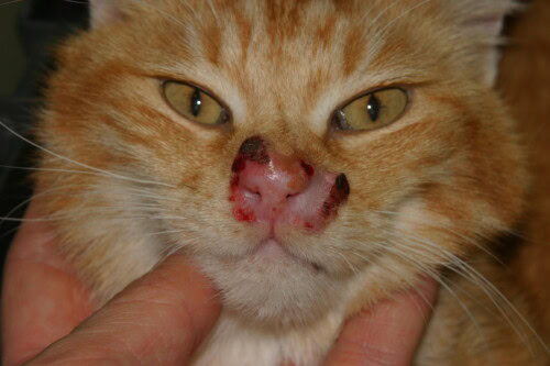
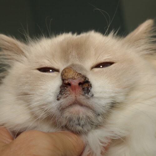
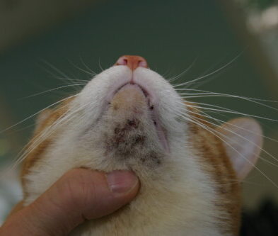
Erosive and crusting facial dermatoses in cats represent a major diagnostic challenge in veterinary practice. Their high prevalence and the multiplicity of possible etiologies require a methodical and rigorous approach. At the last World Congress of Veterinary Dermatology held in Boston, our colleague Petra Bizikova had the opportunity to review their causes and diagnostic approach.
The cat’s face, a privileged interface with the environment, presents anatomical and physiological peculiarities that influence the clinical presentation of these dermatoses. Understanding the origin of facial erosions and crusts requires a thorough knowledge of the underlying pathogenetic mechanisms and a structured diagnostic approach, allowing for appropriate treatment and significantly improving the prognosis.
Physiology and peculiarities of feline facial skin
It has several characteristics that distinguish it from other body regions and influence the presentation of dermatoses in this area. The facial epidermis, relatively thin, comprises 3 to 5 layers of keratinocytes with a thinner stratum corneum than on the rest of the body. This thinness makes it particularly vulnerable to trauma and infections. The underlying dermis is richly vascularized and innervated, explaining the often spectacular clinical expression of pathological processes.
The density of adnexal structures is a remarkable characteristic of the feline face. Hair follicles and their associated glands are particularly numerous, especially on the lips, chin, and cheeks. Sebaceous glands sometimes form specific glandular complexes at the labial commissures and chin, predisposing these areas to certain dermatoses.
Immunologically, facial skin is a particularly active site. The high concentration of Langerhans cells, the main cutaneous antigen-presenting cells, promotes localized immune reactions and partially explains the predisposition of this region to immune-mediated dermatoses. The composition of the resident flora, dominated by coagulase-positive staphylococci and Malassezia, also influences susceptibility to secondary infections.
The intense grooming behavior characteristic of felines plays a major role in the semiology of facial dermatoses. This behavior can not only aggravate pre-existing lesions but also significantly alter their clinical appearance, thus complicating the identification of primary lesions. Since cats use both their rough tongue and their claws during grooming, they can create excoriation lesions that mimic certain primary dermatoses.
Semiology and classification of erosive and crusting facial lesions
Definitions and terminology
Accurate evaluation of dermatological lesions requires standardized terminology for objective and reproducible description.
An erosion corresponds to a loss of skin substance limited to the epidermis, not reaching the dermis. It usually heals without scarring and clinically appears as a moist, depressed, pink to red area. Erosions can result from the rupture of vesicles or pustules, or be secondary to self-trauma.
An ulcer differs from an erosion by a deeper loss of substance, reaching the dermis or underlying tissues. Its healing is usually accompanied by a scar. Ulcers can be deep, hemorrhagic, and painful, with sharp or irregular edges, infiltrated or inflammatory depending on the etiology.
Crusts are solid formations resulting from the desiccation of serous, purulent, or hemorrhagic exudates. They represent secondary lesions often covering underlying erosions or ulcers. Their appearance varies depending on their composition and the age of the lesions: yellowish for serous or purulent exudates, brownish for hemorrhagic exudates.
Primary versus secondary lesions
The distinction between primary lesions (appearing spontaneously) and secondary lesions (resulting from the evolution of a primary lesion or trauma) is fundamental in the diagnostic approach. Crusts and erosions are generally secondary lesions whose origin must be sought. Identifying underlying or prior primary lesions greatly helps in guiding the differential diagnosis.
In cats, observing primary lesions is often complicated by intense grooming, which can quickly alter them. Vesicles and pustules, for example, are particularly fragile and transient in this species, rapidly progressing to erosions and crusts.
Topography and lesion distribution
The precise distribution of facial lesions is a major semiological element guiding the differential diagnosis. Several distribution patterns can be distinguished:
Isolated focal lesions are more indicative of localized neoplastic, traumatic, or infectious processes, while multifocal lesions rather suggest immune-mediated dermatoses, allergic dermatoses, or certain systemic infections.
The preferential location of lesions also provides valuable diagnostic clues:
- Periocular lesions suggest feline herpesvirus infection, Malassezia dermatitis, or certain forms of eosinophilic granuloma complex.
- (Peri)nasal lesions favor pemphigus foliaceus, cutaneous lupus erythematosus, certain forms of dermatophytosis, and squamous cell carcinomas.
- Periauricular lesions are observed in complicated ear mites, certain dermatophytoses, and autoimmune dermatoses.
- Labial and chin lesions suggest chin furunculosis or feline acne.
Lesion symmetry also constitutes an important guiding element. Bilaterally symmetrical lesions are more suggestive of allergic or autoimmune dermatoses, while asymmetry is more indicative of infectious or neoplastic causes.
Evolutionary and clinical characteristics of lesions
The temporal evolution of lesions represents a major diagnostic criterion. Lesions of sudden onset are more indicative of acute infectious causes, allergic reactions, or trauma, while chronic, progressive lesions tend to suggest neoplastic processes or autoimmune dermatoses.
The evaluation of pruritus and pain associated with facial lesions is essential in the diagnostic approach:
- Intense pruritus points to allergic, parasitic, or certain fungal infections.
- Absence of pruritus is more characteristic of neoplastic or autoimmune processes.
- Marked pain may suggest deep ulcerations, secondary bacterial infections, or certain forms of vasculitis.
The macroscopic appearance of crusts also provides valuable diagnostic clues:
- Yellowish crusts suggest pyogenic bacterial infections.
- Thick, adherent, grayish crusts are indicative of dermatophytoses or autoimmune dermatoses like pemphigus foliaceus.
- Brownish or hemorrhagic crusts suggest trauma, vasculitis, or certain ulcerated neoplasms.
Etiology of erosive and crusting facial dermatoses
Infectious dermatoses
Viral infections
Feline herpesvirus (FHV-1) is a common cause of facial dermatosis in cats, particularly in young animals and cats housed in groups. Lesions, usually located around the eyes and nose, begin as vesicles that rapidly evolve into erosions and then crusts. The characteristic “butterfly wing” distribution on the bridge of the nose and nasal planum is relatively specific. Associated conjunctivitis and respiratory symptoms are important guiding elements. The relapsing course, exacerbated by stress or immunosuppression, is typical of this condition. A history of respiratory infection episodes or recent corticosteroid administration often provides revealing anamnestic information.
Feline calicivirus can also cause ulcerative facial lesions, usually associated with oral ulcers and systemic signs (fever, anorexia). Facial lesions primarily involve the nasal philtrum. The virulent systemic form of calicivirus (VS-FCV) can cause severe skin lesions, including facial and extremity ulcerations, associated with vasculitis and marked facial edema.
Feline poxvirus (Cowpox), though rare, typically affects hunting cats with outdoor access. The primary lesion, often located on the head, neck, or forelimbs, presents as a papule or nodule that ulcerates, followed by disseminated similar lesions. General signs (fever, lethargy) frequently accompany this infection. It is a potential zoonosis requiring precautions when handling affected animals.
Feline retroviruses (FeLV, FIV), without directly causing characteristic skin lesions, can promote the development of facial dermatoses through the immunosuppression they induce, facilitating opportunistic infections or the development of certain skin neoplasms.
Feline Herpesvirus
Bacterial infections
Facial pyoderma is common in cats, but almost always secondary to trauma, underlying dermatoses, or local immune deficiencies. Staphylococcus pseudintermedius and S. aureus are the most commonly isolated agents. Clinically, pustules are observed that rapidly evolve into erosions covered with yellowish crusts. Purulent exudate and a favorable response to antibiotics are characteristic. Cytology typically reveals degenerated neutrophils containing intracellular bacteria (cocci).
Chin furunculosis, a particular form of deep pyoderma, manifests as inflammatory papules, pustules, and nodules on the chin, which can evolve into fistulas and hemorrhagic crusts. This condition is thought to be related to an abnormality of hair follicles and sebaceous glands, favoring bacterial proliferation.
Atypical mycobacterial infections (especially Mycobacterium fortuitum and M. chelonae) can cause nodules that ulcerate and become covered with crusts, primarily on the faces of outdoor cats. These infections are often linked to contamination from contaminated scratches or bites. Lesions can form draining tracts, unlike neoplastic processes that ulcerate but generally do not drain.
Fungal infections
Facial dermatophytosis, mainly caused by Microsporum canis, is a common cause of crusting lesions in cats. Classic circular alopecia lesions can be accompanied by erosions and crusts, particularly in young animals, Persian cats, or immunocompromised individuals. The clinical presentation can be extremely variable, including alopecia, scales, erythema, papules, crusts (including miliary), and sometimes inflammatory nodules (kerions). The infection is generally not very pruritic unless there is a co-infection or concomitant allergy. Wood’s lamp fluorescence (positive in 50% of M. canis cases) and direct mycological examination are first-level supplementary examinations.
Malassezia infections can also cause erythematous facial dermatitis with a characteristic brownish exudate, particularly in the periocular regions, chin (acne-like appearance), and facial folds. These infections are generally secondary to an allergy, a keratinization disorder, or a systemic disease. Clinical signs include erythema, brownish to blackish greasy scales or crusts, alopecia, foul odor, and variable pruritus. Diagnosis is based on cytology revealing characteristic “peanut” or “snowman” shaped yeasts.
More rarely, systemic mycoses (cryptococcosis, sporotrichosis, histoplasmosis) can manifest as nodular facial lesions that ulcerate and become covered with crusts. These conditions should be suspected in cats living in endemic areas or presenting underlying immunosuppression.
Parasitic dermatoses
Feline demodicosis, caused by Demodex cati (follicular mite) or D. gatoi (surface mite), can cause erythematous, scaly, and crusting facial lesions, sometimes accompanied by alopecia. The localized form typically manifests as erythema, alopecia, scales, and crusts on the head and neck. Unlike dogs, generalized feline demodicosis is generally associated with underlying immunosuppressive conditions (FIV, FeLV, diabetes, neoplasia) or immunomodulatory treatments. D. gatoi, unlike D. cati, is contagious and often responsible for intense pruritus that can mimic allergic dermatitis. Diagnosis is based on deep skin scrapings for D. cati or superficial scrapings for D. gatoi, the latter sometimes being difficult to detect due to intense grooming.
Notoedric mange (Notoedres cati), although relatively rare, is highly contagious and associated with intense pruritus. Initial lesions are thick crusts localized on the face and the medial edge of the ear pinnae, which can then spread. Diagnosis is based on identification of mites by superficial skin scrapings, which is generally easy. This parasitosis represents a potential zoonosis.
Ear mites (Otodectes cynotis), common in young cats, primarily cause external otitis characterized by black, dry cerumen. Intense pruritus can lead to secondary peri-auricular and facial excoriations and crusts. Diagnosis is made by otoscopy and microscopic examination of the cerumen.
Cheyletiellosis (Cheyletiella blakei), nicknamed “walking dandruff,” is typically characterized by the presence of abundant scales on the back, but can also affect the face and neck. Associated pruritus is variable. This parasitosis is contagious and potentially zoonotic. Diagnosis relies on superficial scrapings, adhesive tape tests, or fine combing.
Trombiculosis (chiggers), a seasonal infestation (late summer/fall) by Neotrombicula autumnalis larvae, can cause erythematous papules that evolve into crusts, primarily localized on the head, ears, limbs, and ventral areas. Associated pruritus is generally intense. Diagnosis is established by direct observation of orange-colored mites grouped on the affected areas.
Immune-mediated dermatoses
Autoimmune dermatoses
Pemphigus foliaceus is the most common autoimmune dermatosis in cats. It is characterized by pustules that rapidly evolve into erosions and yellowish crusts, preferentially located on the face (nose, around the eyes, ear pinnae). Nail folds are frequently affected, developing paronychia with caseous exudate. Other possible locations include paw pads and nipples. Pruritus is variable, and general signs (lethargy, fever, anorexia) may be present. The disease can be idiopathic or induced by certain medications. Cytological diagnosis reveals neutrophils, sometimes eosinophils, and characteristic acantholytic keratinocytes (although these can also be observed in bacterial or fungal infections). Diagnostic confirmation relies on histopathology showing subcorneal or intrafollicular pustules with acanthocytes, in the absence of microorganisms.
Pemphigus foliaceus
Cutaneous lupus erythematosus, rare in cats, typically manifests as symmetrical lesions primarily affecting the nasal planum. One observes a loss of normal cobblestone texture, depigmentation, erythema, scales, erosions, crusts, and sometimes ulceration. The involvement can extend to the periocular skin, ear pinnae, lips, and genitals. These lesions are generally non-pruritic. Diagnosis is based on histopathology revealing an interface dermatitis with basal cell apoptosis.
Systemic lupus erythematosus, even rarer, may include similar facial cutaneous manifestations, associated with systemic signs (polyarthritis, glomerulonephritis, hemolytic anemia).
Arthropod hypersensitivity reactions
Hypersensitivity to mosquito bites can cause papulo-crusting lesions on hairless areas (ear pinna, nasal planum, bridge of the nose), evolving into erosions due to scratching. This condition shows marked seasonality in temperate regions (warm months) and primarily affects outdoor cats. Cytological examination typically reveals marked eosinophilic inflammation, and the anamnesis often reveals recurrent episodes each summer.
Flea bite hypersensitivity can also induce erosive and crusting facial lesions, although the preferential locations are the dorsolumbar region, tail base, and ventral abdomen. Facial lesions usually result from self-trauma secondary to intense pruritus. This is the most common allergy in cats. Diagnosis is suggested by clinical signs, the possible presence of fleas or their droppings (often absent due to intense grooming), and the response to strict antiparasitic control.
Allergic dermatoses
Feline atopic dermatitis, now called feline atopic skin syndrome (FASS), can manifest as erythematous, erosive, and crusting facial lesions, particularly in the periocular area and ear pinnae. The associated pruritus leads to excoriations and secondary ulcerations. This allergy to aeroallergens (dust mites, pollens, molds) often presents with several reaction patterns: cervicofacial pruritus with excoriations, miliary dermatitis, self-induced alopecia, or lesions of the eosinophilic granuloma complex. It can be seasonal or non-seasonal depending on the allergens involved. Its diagnosis is a diagnosis of exclusion, after ruling out parasitoses, infections, FAD, and food allergy. Allergy tests (intradermal tests, serology) can identify the allergens involved for specific immunotherapy, but do not diagnose this condition by themselves.
Food allergy (or cutaneous adverse food reaction, CAFR) presents clinical manifestations often indistinguishable from atopic dermatitis. Facial lesions are common, with intense pruritus, erosions, and crusts affecting the head, neck, and ears. This allergy is generally not seasonal and can occur at any age, although it is more frequent in young adults or middle-aged cats. Its diagnosis relies on a strict elimination diet for 8-12 weeks, followed by a challenge confirming the food origin by the reappearance of clinical signs.
Cutaneous drug reactions can also cause erosive and crusting facial lesions, sometimes associated with systemic manifestations. Antibiotics (especially penicillins and sulfonamides), antiparasitics, and some antifungals are the most frequently implicated.
Cutaneous neoplasms
Epithelial tumors
Squamous cell carcinoma is the most common malignant skin tumor in cats, preferentially affecting poorly pigmented areas exposed to the sun (nasal planum, ear pinnae, eyelids). Lesions classically evolve from an erythematous plaque to a crusting ulceration and then to an infiltrating nodule. Crusting and ulcerative lesions can sometimes resemble other conditions such as feline herpesvirus, mosquito bite hypersensitivity, or even pemphigus foliaceus. This diagnostic confusion is particularly common in early stages, before the mass becomes obvious. Chronic exposure to ultraviolet rays is the main etiological factor, explaining the increased prevalence in white or light-colored cats. Diagnosis relies on histopathology, revealing a proliferation of atypical keratinocytes invading the dermis.
Squamous cell carcinoma in situ (BISC, also known as Bowen’s disease or viral plaques) is associated with feline papillomavirus and potentially retroviruses (FIV/FeLV). This neoplasm often affects older cats (>10 years) and presents as multifocal, pigmented or non-pigmented, crusting, hyperkeratotic, sometimes ulcerated plaques, which can affect the head and face. Diagnosis relies on histopathological examination.
Cutaneous mast cell tumor
Feline cutaneous mast cell tumor generally presents as solitary or multiple nodules that can ulcerate and become covered with crusts. Facial localization is less common than on the trunk or limbs, but still possible. Unlike in dogs, feline cutaneous mast cell tumors generally exhibit benign behavior. Cytological diagnosis reveals a homogeneous population of mast cells containing metachromatic granules with appropriate stains. Surgical excision is usually the treatment of choice.
Cutaneous lymphoma
Epitheliotropic cutaneous lymphoma (mycosis fungoides) can manifest as erythematous, erosive, and crusting lesions affecting the face, among other areas. The evolution is classically progressive, starting with an erythematous phase (patch stage), evolving to an infiltration phase (plaque stage), and then a tumor phase. Diagnosis relies on histopathology supplemented by immunohistochemistry, allowing characterization of the tumor immunophenotype.
Paraneoplastic syndromes
Exfoliative dermatitis associated with thymoma is a rare paraneoplastic syndrome affecting middle-aged or older cats. This non-pruritic, severely flaky, erythematous, crusting, and alopecic dermatitis often begins on the head and neck before becoming generalized. Cutaneous signs usually precede the clinical manifestations of the underlying thymoma. Diagnosis relies on cutaneous histopathology associated with thoracic imaging (radiography, ultrasound) to detect the thymic mass. Resolution of cutaneous signs after thymoma excision confirms the paraneoplastic nature of the syndrome.
Keratinization disorders
Feline idiopathic facial dermatitis in Persians is a well-known but poorly understood condition specific to Persian and Himalayan cats. It generally appears before one year of age and is characterized by severe inflammation with accumulation of dark seborrheic material around the eyelids, lips, chin, and sometimes ear canals. This material adheres to skin folds, giving a characteristic “dirty face” appearance. The accumulation of this debris promotes secondary bacterial superinfections, aggravating the clinical picture with the formation of true crusts, exudative, erosive, and ulcerative lesions, accompanied by variable pruritus, pain, and alopecia. Treatment is symptomatic and generally lifelong, including antiseborrheic and antimicrobial topical products, sometimes associated with oral steroids, cyclosporine, or topical tacrolimus.
Sebaceous gland dysplasia is a congenital condition characterized by early manifestations, as early as the first months of life. Clinically similar to idiopathic facial dermatitis, it differs in its extension beyond the face, with generalized lesions affecting the rest of the body. Facial seborrheic accumulation is accompanied by follicular casts, poor coat quality with hair thinning and alopecia. A recent genetic study identified a missense mutation in the SOAT1 gene encoding sterol O-acetyltransferase 1, an enzyme responsible for the formation of cholesterol esters necessary for the normal composition of sebum and meibum. This mutation likely explains the abnormalities in sebaceous production and hair growth disorders. No specific treatment is currently available for this genetic condition.
Nasal hyperkeratosis of Bengal cats is an emerging entity, probably hereditary and also reported in Egyptian Maus. It usually appears during the first year of life and is characterized by significant hyperkeratosis of the nasal planum that can progress to fissures and ulcerations. Lesions remain strictly limited to the nasal planum and cause variable discomfort. Spontaneous improvements have been reported in some cases, while others seem to respond to topical tacrolimus. Genetic research is underway to identify the responsible gene(s).
Other causes
Feline acne is characterized by the presence of comedones, papules, and pustules on the chin, which can progress to furuncles and fistulas in severe forms. Crusting results from the drying of inflammatory exudate. The etiology involves follicular hyperkeratosis and excessive sebum production, potentially exacerbated by genetic and environmental factors.
Behavioral dermatoses resulting from self-trauma (excessive licking, scratching) related to stress, anxiety, or frustration, can cause alopecia, erosion, ulceration, and crusting lesions, often in accessible areas like the face and neck. These dermatoses particularly affect cats living exclusively indoors in an insufficiently enriched environment. Their diagnosis is a diagnosis of exclusion, after having ruled out all medical causes.
Trauma and burns can also be responsible for erosive and crusting facial lesions that are sometimes difficult to distinguish from primary dermatoses. The absence of specific lesions and medical history generally guide the diagnosis.
Diagnostic approach to erosive and crusting facial dermatoses
Anamnesis and clinical examination
Key anamnestic elements
The investigation of facial dermatoses begins with a meticulous anamnesis, a true cornerstone of the diagnostic process. The following information should be systematically collected:
- Lifestyle: outdoor access, cohabitation with other animals, environment (recent changes)
- Precise chronology of lesions: date of onset, evolution (acute or progressive), possible seasonal variations, response to previous treatments
- Pruritus: presence or absence, intensity, appearance before or after visible skin lesions
- Associated systemic signs: appetite or weight disorders, behavioral changes, concomitant respiratory or digestive symptoms
- Medical history: known chronic pathologies, current drug treatments (especially corticosteroids or immunosuppressants)
- Dermatological history: previous similar episodes, recurrent dermatoses
- Diet: type, recent changes, food supplements
- Contagiousness: affection of other animals or people in contact
This information significantly guides the differential diagnosis and allows for effective planning of appropriate complementary examinations.
Dermatological examination
The dermatological examination must be methodical and exhaustive. It begins with a distant observation allowing evaluation of the general distribution of lesions, followed by a close examination:
- Precise description of lesions: type (primary and secondary), size, distribution, symmetry
- Careful search for primary lesions often masked by secondary lesions
- Evaluation of extension: involvement limited to the face or presence of lesions on other body regions
- Examination of mucous membranes and mucocutaneous junctions: ulcerations, depigmentation
- Palpation of lesions: consistency, adherence to deep planes, sensitivity or pain
- Examination of adnexa: coat quality, presence of alopecia, condition of claws and paw pads
The use of a tangential light source facilitates the visualization of discrete lesions, particularly superficial erosions. Wood’s lamp examination, performed in the dark after adequate preheating of the lamp, can reveal the characteristic greenish fluorescence of certain strains of Microsporum canis.
General clinical examination
The general clinical examination should never be neglected, as many facial dermatoses fit into a broader pathological context:
- Lymph node examination: regional lymphadenopathy may suggest an infectious or neoplastic process.
- Oral cavity examination: the presence of ulcerations, stomatitis, or gingivitis is particularly important in viral infections or certain autoimmune diseases.
- Cardiopulmonary auscultation: search for associated respiratory signs, especially in herpesvirus infection.
- Temperature measurement: hyperthermia may point to an infectious or systemic inflammatory cause.
- Ophthalmological examination: conjunctivitis, keratitis, uveitis that may be associated with certain facial dermatoses of viral or immune origin.
First-line complementary examinations
Cutaneous cytology
Cutaneous cytology is a simple, rapid, and inexpensive complementary examination, often providing valuable information for etiological diagnosis. Sampling techniques vary depending on the type of lesion:
- Direct impression smear for exudative lesions.
- Scraping and then spreading for previously moistened crusts.
- Fine-needle aspiration for nodular lesions.
After rapid staining (Diff-Quik® type), microscopic examination looks for:
- Inflammatory cells: predominance of neutrophils indicates bacterial infection or pemphigus foliaceus; predominance of eosinophils suggests allergy, parasitism, eosinophilic granuloma complex, herpesvirus infection, or sometimes pemphigus foliaceus.
- Infectious agents: bacteria (cocci, bacilli), yeasts (Malassezia recognizable by their “peanut” or “snowman” shape), fungal spores.
- Acantholytic cells: rounded keratinocytes detached from each other, highly suggestive of pemphigus foliaceus, although not pathognomonic.
- Neoplastic cells: cytological atypia, monomorphic populations.
The discovery of acantholytic keratinocytes is a critical point in cytological interpretation. Although highly suggestive of pemphigus foliaceus, this anomaly can also be observed in bacterial (staphylococcal) or fungal (dermatophyte) infections. Before concluding pemphigus and considering a biopsy or immunosuppressive treatment that could be detrimental in the case of underlying infection, it is imperative to rigorously rule out pyoderma and dermatophytosis by appropriate complementary examinations.
Skin scrapings
Skin scrapings are a fundamental diagnostic technique for the search for ectoparasites, particularly Demodex and Notoedres. Two types of scrapings are performed:
- Superficial scraping: performed by gently scraping the skin surface with a dull blade and mineral oil. It aims to collect surface mites such as Notoedres cati (prefer the edge of the ears), Cheyletiella, Demodex gatoi (sometimes difficult to detect), and Otodectes (on the periauricular skin).
- Deep scraping: necessary to search for follicular mites such as Demodex cati. It requires firmly pinching a skin fold and scraping until capillary bleeding occurs to access follicular contents.
Direct microscopic examination of the scraping product is carried out between a slide and a coverslip, possibly after clarification with 10% potassium hydroxide.
Fungal culture
Fungal culture on Sabouraud’s medium (or DTM, Dermatophyte Test Medium) is the reference examination for the diagnosis of dermatophytosis. Samples are taken by brushing with a sterile brush or by plucking hairs from the periphery of the lesions. Incubation is carried out at 25-30°C for three weeks with regular observation. On DTM, interpretation is based on the joint observation of a color change of the medium to red (alkalinization due to dermatophyte metabolism) and the growth of a white or buff colony. Microscopic identification of macroconidia is necessary to confirm the species of dermatophyte involved.
Direct microscopy of hairs after clarification with 10-30% KOH can sometimes rapidly visualize fungal arthrospores surrounding hair shafts, providing a quick presumptive diagnosis.
Second-line complementary examinations
Skin biopsies
Skin biopsy often represents the examination of choice for establishing a definitive diagnosis, particularly in cases of immune-mediated, neoplastic, or refractory dermatoses to empirical treatments. The quality of the sample strongly conditions the diagnostic value of the histopathological examination:
-
Sampling technique:
- Punch biopsy (4-6 mm diameter) or excisional biopsy with a scalpel in some cases
- Sampling of recent, untreated lesions, avoiding chronic, altered lesions
- Inclusion of the junction between healthy and lesional tissue
- Multiple biopsies (3-5 sites) to increase diagnostic sensitivity
- Specific technique for vesicular or erosive lesions: for intact vesicles, sample centered on the vesicle surrounded by a few millimeters of healthy skin; for erosions, sample at the margin between eroded and intact skin
-
Practical considerations:
- Prior treatment of secondary infections before biopsy to avoid diagnostic artifacts
- Withdrawal of immunosuppressants for a sufficient period to allow characteristic lesion expression
- Preservation of crusts that may contain important diagnostic elements (acantholytic cells, fungal hyphae)
- Appropriate orientation of samples to facilitate histological preparation, particularly for margin biopsies
- Detailed communication of clinical information to the pathologist
Histopathological interpretation should be entrusted to a pathologist experienced in veterinary dermatopathology. In some cases, special stains may be necessary to highlight infectious agents (PAS for fungi, Ziehl-Neelsen for mycobacteria) or specific tissue characteristics.
Immunodiagnostic techniques
Immunodiagnostic techniques are particularly useful for confirming autoimmune dermatoses:
- Direct immunofluorescence (DIF) allows detection of immunoglobulin and complement deposits in the skin. In pemphigus foliaceus, intercellular deposits of IgG and C3 are observed in the superficial epidermis. In lupus erythematosus, linear or granular deposits of immunoglobulins and C3 are found at the dermo-epidermal junction.
- Indirect immunofluorescence (IIF) searches for circulating autoantibodies in serum. This technique has lower sensitivity than DIF in feline autoimmune dermatoses, but can confirm some cases of pemphigus foliaceus.
- Specific ELISA tests have been developed for the detection of anti-desmoglein 1 antibodies in feline pemphigus foliaceus, but their availability remains limited in clinical practice.
These examinations require specific samples: frozen biopsies or those fixed in Michel’s solution for DIF, and blood samples for IIF and ELISA.
Allergy tests
Allergy tests may be indicated in cases of suspected allergic facial dermatoses:
- Intradermal tests: intradermal injection of standardized allergen dilutions, with reading after 15-30 minutes. These tests have limited specificity in cats and generally require sedation.
- Serological tests: search for specific IgE to environmental allergens in serum. These tests are easier to perform but their interpretation remains delicate due to frequent false positive results.
- Elimination-challenge diet: constitutes the reference method for diagnosing food allergy. It consists of a strict hypoallergenic diet for 8-10 weeks, followed by a gradual reintroduction of suspected foods.
These tests cannot directly diagnose feline atopic skin syndrome, which remains a diagnosis of exclusion. Their primary aim is to identify the allergens involved to guide allergen avoidance or specific immunotherapy.
Specific complementary examinations
Certain facial dermatoses require specific diagnostic tests:
- PCR (Polymerase Chain Reaction): particularly useful for the detection of feline herpesvirus (FHV-1) on conjunctival swabs, corneal scrapings, or skin biopsies. This technique has high sensitivity and specificity but cannot distinguish between active infection and latent carriage.
- Bacterial cultures and antibiograms: indicated in recurrent pyoderma or those refractory to first-line treatments. Samples should be taken before any antibiotic therapy, by biopsy or aspiration of closed abscesses to avoid contamination.
- FIV/FeLV serology: recommended for cats with atypical, multifocal, chronic, or generalized facial dermatoses, especially in cases of associated systemic signs or suspected immunosuppression.
- Imaging examinations: rarely indicated for isolated facial dermatoses, but may be necessary in certain specific contexts such as the search for an underlying thymoma (radiography, thoracic ultrasound) in case of suspected paraneoplastic exfoliative dermatitis.
Diagnostic algorithm
The diagnostic approach to erosive and crusting facial dermatoses can be systematized according to a decision-making algorithm based on clinical characteristics and the results of complementary examinations.
Initial assessment
The first step is to determine whether the facial dermatosis is part of a broader clinical picture or remains strictly localized to the face:
- Generalized dermatosis with facial involvement: points more towards systemic causes, including allergic, autoimmune, or generalized parasitic dermatoses.
- Strictly facial dermatosis: rather suggests local causes, such as localized infections, neoplasms, actinic dermatoses, or keratinization disorders.
Classification based on the presence of pruritus
Pruritus is a major guiding element:
- Pruritic dermatosis: mainly suggests parasitic causes (notoedric mange, Demodex gatoi, trombiculosis), allergic causes (atopic dermatitis, food allergy) or certain infections (dermatophytosis, Malassezia dermatitis).
- Non-pruritic dermatosis: points more towards neoplastic, autoimmune, uncomplicated viral, or metabolic causes.
It should be noted, however, that the intensity of pruritus can be modified by previous treatments (especially corticosteroids) or masked by painful lesions.
Orientation according to lesion topography
The precise distribution of facial lesions provides important diagnostic clues:
- Symmetrical bilateral lesions: characteristic of immune-mediated dermatoses (pemphigus foliaceus, lupus erythematosus) and certain allergic dermatoses.
- Asymmetrical or unilateral lesions: more suggestive of localized infectious, traumatic, or neoplastic causes.
- Peri-orificial lesions: preferential involvement of mucocutaneous junctions (nostrils, lips, eyelids) is typical of pemphigus foliaceus and certain forms of cutaneous lupus erythematosus.
Structured diagnostic approach
A sequential and logical approach is indispensable for navigating among the numerous differential diagnoses:
- Step 1: Detailed anamnesis and clinical examination (signalment, lifestyle, lesion history, pruritus, systemic signs)
- Step 2: First-line complementary examinations, performed jointly
- Cytology (mandatory)
- Skin scrapings (superficial and/or deep depending on clinical suspicion)
- Wood’s lamp examination
- Fungal culture
- Search for ectoparasites (fine combing, brushing)
- Step 3: Initial therapeutic trials based on previous results
- Specific antiparasitic treatment if parasites identified
- Appropriate antimicrobial treatment if bacterial or fungal infection detected
- Strict flea control trial (8-9 weeks) if allergy suspected
- Elimination diet (8-12 weeks) if food allergy remains suspected after exclusion or treatment of other causes
- Step 4: Re-evaluation and second-line complementary examinations
- Skin biopsies for histopathology if immune-mediated, neoplastic, viral, or atypical inflammatory dermatosis suspected
- Allergy tests if feline atopic skin syndrome suspected after exclusion of other causes
- Serological tests (FIV/FeLV) if immunosuppression suspected
- Viral PCR if herpesvirus or calicivirus infection suspected
- Bacterial culture with antibiogram if refractory pyoderma
This diagnostic approach is fundamentally iterative. The response – or lack thereof – to initial treatments is itself a valuable diagnostic element. The failure of a well-conducted empirical treatment should lead to reconsideration of diagnostic hypotheses and progression to more in-depth investigations.
Predisposing factors and prognostic considerations
Age is an important epidemiological factor in the diagnostic approach to facial dermatoses:
-
Kittens and young cats (<1-3 years): Are more frequently affected by dermatophytosis, ectoparasitosis (Otodectes, Cheyletiella, Notoedres), certain viral infections (papillomavirus, herpesvirus). Allergies often begin at a young age, typically before 3-4 years. Idiopathic facial dermatitis of Persians and nasal hyperkeratosis of Bengals generally appear before one year of age. Congenital problems like sebaceous gland dysplasia manifest early.
-
Adult and older cats: Pemphigus foliaceus occurs on average around 5 years of age, but with a wide age range. Neoplasms and paraneoplastic syndromes are more frequent in older cats (squamous cell carcinoma, carcinoma in situ, thymoma-associated dermatitis). Herpesvirus infection can occur at any age, but reactivations are more frequent in older or stressed cats. Systemic diseases predisposing to secondary infections (diabetes, hyperadrenocorticism, retroviruses) are more common in adults and seniors.
Breed predispositions
Certain breeds have predispositions to developing specific facial dermatoses:
-
Persians and Himalayans: Predisposed to dermatophytosis, idiopathic facial dermatitis, primary seborrhea. Pemphigus foliaceus has also been reported.
-
Abyssinians: Possible predisposition to feline atopic skin syndrome.
-
Devon Rex: Predisposed to Malassezia proliferation and associated dermatitis. Predisposition to feline atopic skin syndrome reported.
-
Siamese: Possible predisposition to feline atopic skin syndrome.
-
Brachycephalic breeds: Problems related to facial folds favor bacterial and fungal superinfections.
-
Bengals and Egyptian Maus: Hereditary nasal hyperkeratosis.
-
Sphynx: Frequent carriers of Malassezia.
However, it should be emphasized that breed predispositions are generally less well-defined in cats than in dogs and should not replace a complete diagnostic approach. Many conditions affect European or mixed-breed cats equally.
Influence of lifestyle and environment
The cat’s lifestyle and environment significantly influence the risk of developing certain facial dermatoses:
-
Outdoor access: Increases the risk of poxvirus (hunting rodents), trauma and abscesses, parasitic infestations (fleas, ticks, chiggers), and potentially dermatophytosis.
-
Group living (shelters, catteries, multi-cat households): Increases the risk of contagious diseases such as dermatophytosis, cheyletiellosis, notoedric mange, ear mites, Demodex gatoi, and viral infections (FCV, FHV-1).
-
Diet: Dietary history is crucial for the diagnosis of food allergy. Inadequate nutrition can affect skin health.
-
Stress and anxiety: Can trigger reactivation of herpesvirus and contribute to behavioral dermatoses.
-
Immunosuppression: An underlying disease (FIV, FeLV, diabetes, hyperadrenocorticism, neoplasia) or certain treatments (glucocorticoids, chemotherapy) increase the risk of opportunistic infections such as generalized Demodicosis due to D. cati, Malassezia proliferation, severe or atypical dermatophytosis, and deep pyoderma.
Immunosuppression, particularly linked to FIV or FeLV infections, is a recurrent risk factor for several distinct facial dermatoses. This constant association highlights the paramount importance of considering and testing retroviral status in cats presenting with severe, chronic, unusual, or generalized facial dermatoses. The diagnostic approach is not limited to treating the skin lesion but also involves identifying potential underlying serious diseases.
Prognostic considerations
The prognosis of erosive and crusting facial dermatoses depends on multiple factors:
-
Nature of the underlying condition: Infectious and parasitic causes generally have a good prognosis with appropriate treatment. Allergic dermatoses can be controlled but rarely definitively cured. Neoplasms have a variable prognosis depending on their nature, extent, and available therapeutic options.
-
Early diagnosis: A rapid diagnosis generally allows for more effective management and limits secondary complications.
-
Presence of concomitant diseases: Comorbidities, particularly underlying immunosuppressive diseases, can worsen the prognosis and complicate therapeutic management.
-
Treatment adherence: The often chronic nature of facial dermatoses frequently requires prolonged or lifelong treatments, whose adherence determines the clinical course.
-
Zoonotic potential: Certain conditions such as dermatophytoses, cheyletiellosis, notoedric mange, or poxvirus present a zoonotic risk requiring special precautions.
Conclusion
Erosive and crusting facial dermatoses in cats represent a heterogeneous group of conditions whose diagnosis constitutes a genuine challenge in clinical practice. The multiplicity of possible causes and the frequent similarity of clinical presentations necessitate a systematic and rigorous diagnostic approach.
An effective diagnostic approach relies on several fundamental pillars: a detailed anamnesis, a thorough clinical examination, first-line complementary examinations (cytology, scrapings, mycological examinations), and, if necessary, second-line investigations (biopsy, specific cultures, allergy tests, serological or molecular biology examinations).
Certain clinical elements prove particularly guiding: intense pruritus strongly suggests a parasitic or allergic origin; specific lesions of the nasal planum may suggest herpesvirus infection, discoid lupus, or mosquito bite hypersensitivity; concomitant involvement of the nail folds is highly suggestive of pemphigus foliaceus; the presence of general signs should prompt investigation for systemic disease or viral infection such as poxvirus.
It is crucial to identify and treat secondary infections (bacterial, Malassezia) which frequently complicate the clinical picture. However, long-term therapeutic success relies on identifying and managing the underlying primary cause. Clear communication with the owner and regular follow-up are essential to adapt treatment and prevent recurrences.
Finally, advances in feline dermatology, both diagnostically and therapeutically, now allow for optimized management of these complex conditions, significantly improving the prognosis and quality of life of affected cats.


