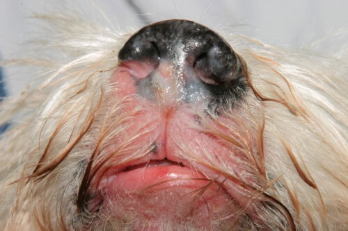
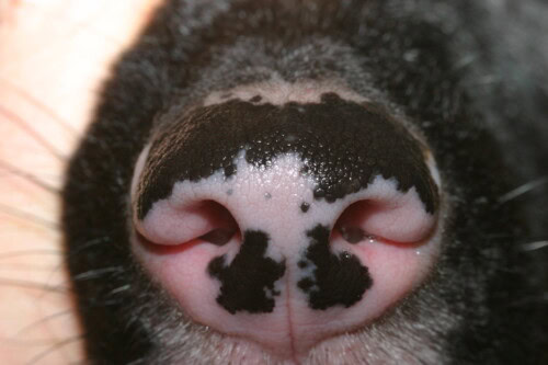
The canine nasal planum, a glabrous and aglandular structure, is subject to a variety of dermatological pathologies. These dermatoses often share similar clinical characteristics, making diagnosis a challenge for the veterinary practitioner.
This article aims to describe the most frequent nasal planum dermatoses in dogs, examining their clinical, diagnostic, and histopathological characteristics, while also addressing their pathophysiology when established. The treatment of these conditions will not be covered in this review.
Classification of Nasal Planum Dermatoses
Dermatoses affecting the nasal planum can be classified according to the type of lesion: crusting and ulcerative, depigmenting, nodular, and hyperkeratotic, bearing in mind that a dermatosis can manifest with several types of manifestations. Another classification is possible based on the underlying pathological process: infectious, allergic, immune-mediated, neoplastic, and miscellaneous/hereditary. This approach facilitates differential diagnosis.
Crusting and Ulcerative Dermatoses
Pemphigus Foliaceus (PF)
Pemphigus foliaceus is the most common immune-mediated dermatosis in dogs. It is characterized by an attack and destruction of desmosomes (intercellular bridges) in the subcorneal layers between keratinocytes, involving IgG autoantibodies targeting desmocollin-1. Although often idiopathic, it can be triggered by drugs or neoplasia. Lesions manifest as large pustules evolving into erosions covered with thick serocellular crusts. Frequently affected sites include the nasal planum, paw pads, ear pinnae, and the haired skin of the face and trunk. Diagnosis relies on a skin biopsy, ideally of an intact pustule. Histopathological examination will reveal large pustules and crusts containing neutrophils and a variable number of eosinophils, with acantholytic keratinocytes. The epidermis is hyperplastic and spongy.
Pemphigus Vulgaris (PV)
Pemphigus vulgaris is a rare autoimmune condition targeting desmosomes in the deeper parts of the epidermis, specifically desmoglein-3. Onset is usually sudden, with ulcers of the mucous membranes and mucocutaneous junctions. Unlike PF, PV is primarily a mucosal disease that can also affect mucocutaneous junctions and, rarely, haired skin. The main lesion is a vesicle or bulla rapidly evolving into ulcers. Diagnosis requires a skin or mucosal biopsy from an intact vesicle, or, failing that, the edges of a recent ulcer. Histopathology will show suprabasal cleavage and acantholysis with pustule or bulla formation.
Mucous Membrane Pemphigoid (MMP)
Mucous membrane pemphigoid is the most common subepidermal autoimmune bullous disease in dogs. It results from autoantibodies targeting components of the basement membrane, including collagen XVII, BP230, and/or laminin 332. The symmetrical lesions consist of fragile bullae and vesicles rapidly evolving into erosions and ulcers. Diagnosis relies on biopsy, revealing subepidermal cleavage above the lamina densa.
Eosinophilic Furunculosis
Facial eosinophilic furunculosis is an acute-onset condition primarily affecting young large-breed dogs. It is characterized by erythematous papules or nodules rapidly evolving into erosions and ulcers with hemorrhagic crusts. The lesions are located on the dorsal muzzle and sometimes extend to the edges of the nasal planum without penetrating it. Cytology and histopathology confirm the diagnosis, which is based on a significant dermal infiltration of eosinophils around adnexal units.
Discoid Lupus Erythematosus (DLE)
Several forms of cutaneous lupus erythematosus (CLE) exist, which are immune-mediated skin diseases sharing histopathological characteristics: a predominantly lymphocytic cytotoxic interface dermatitis. Localized/facial DLE is primarily limited to the nasal planum, but can also affect the nostrils, lips, periorbital skin, and ear pinnae. It most commonly manifests as nasal depigmentation, with loss of the cobblestone architecture and the appearance of crusts and ulcerations. Differential diagnosis between DLE and other conditions such as mucocutaneous pyoderma (MCP) can be difficult and requires a study of the clinical history, lesion topography, and response to antibiotic treatment. Histologically, DLE is characterized by cytotoxic interface dermatitis.
Mucocutaneous Pyoderma (MCP)
Mucocutaneous pyoderma is a bacterial infection of the mucocutaneous junctions, frequently diagnosed in German Shepherds, but not exclusively. It is difficult to distinguish from DLE or MMP. Lesions are symmetrical and consist of erythema, crusts, fissures, and ulcers. Cytology should look for the presence of cocci. Diagnosis relies on response to antibiotic treatment. Histologically, a superficial lymphocytic band and a hyperplastic and variably spongiotic epidermis are observed.
Nasal Arterioplasty
Nasal arteritis refers to a vasculopathy where arteries and arterioles appear to be targeted by the immune system. Two distinct clinical entities have been described: Philtrum Nasal Dermal Arteritis (PNDA) and Nasal Alar Rostrolateral Arterioplasty (NARLA) of German Shepherds. PNDA causes a well-demarcated linear ulcerated fissure on the nasal philtrum. NARLA is characterized by vertical fissures on one or both dorsolateral alar wings. Histologically, the surface is ulcerated secondary to ischemia, and the underlying dermis contains a perivascular infiltrate of inflammatory cells. The deep arteries and arterioles show luminal narrowing.
Depigmenting Dermatoses
Uveodermatologic Syndrome
This is a rare immune-mediated disease in which pigmented tissues are attacked. The condition is characterized by rapid progression of clinical signs, including uveitis and cutaneous depigmentation. It occurs mainly in Akitas and other Arctic breeds. Depigmentation is noticeable on the lips, nasal planum, periorbital skin, and oral mucous membranes. Untreated progression leads to irreversible blindness. Histologically, a superficial dermal infiltration of macrophages containing melanin pigment and loss of epidermal pigmentation are observed.
Cutaneous Epitheliotropic T-Cell Lymphoma
In this dermatosis, neoplastic T-lymphocytes invade the cutaneous and follicular epidermis. It occurs mainly in older dogs, more often in Golden Retrievers. Nasal planum lesions consist of depigmentation, erythema, and ulceration. Other mucous membranes and mucocutaneous junctions may be affected. Skin biopsy is necessary for diagnosis. Histopathology reveals an infiltration of neoplastic T-lymphocytes in the epidermis, forming microaggregates.
Vitiligo
Vitiligo is a depigmenting disorder in which hair and skin gradually whiten due to the destruction of melanocytes. It can occur in all breeds and cause localized or generalized depigmentation. Histopathology shows the absence of melanin pigment in the epidermis.
Nodular Dermatoses
Squamous Cell Carcinoma (SCC)
Squamous cell carcinoma is a common neoplasm of keratinocytes, often associated with chronic sun exposure. On the nasal planum, it can present as generalized or multifocal ulceration, or as eroded plaques or nodules. Cytological examination may reveal atypical epithelial cells, but histopathology is essential for diagnosis.
Hyperkeratotic Dermatoses
Zinc-Responsive Dermatosis
This is a keratinization disorder associated with inherited (syndrome I) or acquired (syndrome II) zinc deficiency. Syndrome I is observed mainly in Arctic breeds, while syndrome II occurs in very young dogs of all breeds. It is characterized by thick hyperkeratotic crusts. Histologically, the stratum corneum is widened by parakeratotic hyperkeratosis.
Hereditary Nasal Parakeratosis of Labrador Retriever
Hereditary nasal parakeratosis of Labrador Retriever is characterized by an accumulation of thick layers of compressed keratin on the dorsal nasal planum. The condition appears to be autosomal recessive. Histologically, diffuse parakeratotic hyperkeratosis that widens the stratum corneum is observed.
Nasodigital Hyperkeratosis
It refers to hyperkeratosis affecting both the nasal planum and paw pads. It can occur in young dogs as a hereditary condition or as an age-related idiopathic change. The nose shows rough keratin accumulations, and the paw pads show fern-like hyperkeratosis. Diagnosis relies on clinical appearance, but biopsy can be considered.
Xeromycteria and Parasympathetic Nose
Hyperkeratosis can also result from extreme dryness of the nasal planum and mucous membranes (xeromycteria). Xeromycteria can be due to loss of parasympathetic signaling from the facial nerve, also leading to neurogenic keratoconjunctivitis sicca. Histopathology may resemble that of nasal hyperkeratosis.
Conclusion
Canine nasal planum dermatoses present a variety of underlying causes. Categorization by lesion type, signalment, and clinical characteristics helps narrow down differential diagnoses. Cytology should be performed to check for bacterial or fungal infection. Biopsy is a powerful diagnostic method for most nasal planum dermatoses. Treating any infection before biopsy and providing complete clinical information increase the chances of obtaining an accurate diagnosis.
Citron L, Brame B, Bradley C. Nasal Planum Dermatoses of the Dog: Clinical Presentations and Diagnostic Approach. Vet Clin Small Anim. 2024;54(6):1257-1278. https://doi.org/10.1016/j.cvsm.2024.11.011

