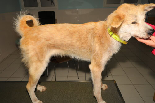
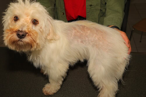
On the occasion of GEDAC’s last annual meeting in Ajaccio in June 2024, our colleague Sébastien VIAUD, DipECVD, gave an overview of this largely unknown dermatosis.
Introduction: A complex and confusing skin disease
Granulomatous sebaceous adenitis (GSA) is an idiopathic inflammatory dermatosis characterized by a progressive and irreversible destruction of the sebaceous glands, which are essential for the production of sebum, a crucial element for skin homeostasis. Although rare, this disease raises persistent questions regarding its etiology and pathophysiology, leading to significant difficulties in diagnosis and therapeutic management.
The impact of GSA on animal welfare is undeniable. Affected dogs may experience unsightly skin lesions, severe itching, and recurrent infectious complications, significantly impairing their quality of life. Furthermore, sebaceous adenitis represents an emotional and financial burden for owners who face a chronic illness that requires constant care and regular veterinary visits. In the most severe cases, euthanasia may be considered as a last resort to end the animal’s suffering.
This conference report aims to provide a comprehensive overview of current knowledge on canine sebaceous adenitis, based on data available in the scientific literature. We will examine epidemiological aspects, pathophysiological hypotheses, clinical manifestations, diagnostic approaches, therapeutic strategies, and research perspectives in this field.
Epidemiology: Prevalence and Risk Factors
Sebaceous adenitis is considered a rare disease in dogs. Its exact prevalence is difficult to estimate due to a lack of large-scale epidemiological studies. However, data collected in various retrospective studies indicate that GSA preferentially affects young or middle-aged adult dogs between 1 and 5 years of age.
Breed Predispositions: Important Genetic Clues
Marked breed-specific predispositions have been observed, strongly suggesting that genetic factors are involved in the etiology of sebaceous adenitis. The most commonly affected dog breeds are:
-
Akita Inu: An autosomal recessive inheritance pattern has been clearly demonstrated in this breed, meaning that affected dogs have inherited two copies of the defective gene, one from each parent.
-
Standard Poodle: Studies have also shown a genetic predisposition for GSA in Standard Poodles, although the mode of transmission has not yet been precisely clarified.
The Akita Inu is the breed most commonly affected by GSA.
Other dog breeds potentially predisposed to sebaceous adenitis have been reported, including:
-
Havanese
-
Lhasa Apso
-
Chow-Chow
-
German Shepherd
-
Samoyed
-
Vizsla
-
Springer Spaniel
But other breeds are also affected.
The observation of a familial aggregation of GSA cases, with multiple puppies from a litter being affected, strengthens the hypothesis of a predominant genetic component in the development of this disease.
No Obvious Sex Predisposition
A significant sex predisposition has not been definitively established in canine sebaceous adenitis. Some studies have suggested a slightly higher prevalence in males, but these observations have not been confirmed by other works. It is therefore likely that both sexes are equally affected.
Pathophysiology: Unravelling the Complex Mechanisms of Sebaceous Gland Destruction
The pathophysiology of canine sebaceous adenitis remains a research area. Despite numerous studies, the exact mechanisms leading to the destruction of sebaceous glands are still unclear. Several hypotheses, often interconnected, have been put forward to explain this complex pathological process:
1. Hypothesis of Hereditary Defect in Sebaceous Gland Development:
This hypothesis suggests that a genetic anomaly, transmitted hereditarily, affects the development and differentiation of sebaceous glands from embryonic or fetal stages. This developmental defect could make the sebaceous glands more susceptible to subsequent damage, leading to their premature destruction.
Arguments for this hypothesis:
-
Marked Breed Predisposition: The existence of strong breed predispositions suggests a genetic basis for the disease.
-
Possible Involvement of Multiple Puppies from a Litter: The familial clustering of GSA cases strengthens the hypothesis of hereditary transmission.
2. Hypothesis of an Autoimmune Disease:
According to this hypothesis, sebaceous adenitis would be an autoimmune disease, in which the dog’s immune system attacks its own sebaceous glands, recognizing them as foreign elements.
Arguments for this hypothesis:
-
Presence of a Lymphocytic Inflammatory Infiltrate Around the Sebaceous Glands: Histopathological examination of skin biopsies shows the presence of T lymphocytes and antigen-presenting dendritic cells around the hair follicles and sebaceous ducts, suggesting an immune reaction against these structures.
-
Efficacy of Certain Immunomodulators used in the treatment of autoimmune diseases. This could suggest an involvement of the immune system in the pathogenesis of the disease.
3. Hypothesis of a Primary Keratinization Disorder:
Some authors suggest that sebaceous adenitis could be a consequence of a primary abnormality of follicular keratinization. Follicular hyperkeratosis, characterized by an excessive accumulation of keratin in the hair follicles, is a histological lesion frequently observed in GSA.
Arguments for this hypothesis:
-
Follicular Hyperkeratosis: The presence of marked follicular hyperkeratosis could clog the sebaceous ducts, preventing normal sebum flow and promoting inflammation and secondary destruction of the sebaceous glands.
-
Efficacy of Retinoids in Some Cases: Retinoids, molecules that modulate the differentiation and proliferation of keratinocytes, have shown some efficacy in treating GSA in dogs in some cases, which could suggest a role of keratinization in the pathogenesis of the disease.
4. Hypothesis of a Lipid Metabolism Anomaly:
It is also possible that a lipid metabolism anomaly, affecting sebum composition or follicular keratinization, could play a role in the development of sebaceous adenitis.
Arguments for this hypothesis:
-
Positive Response to Vitamin A, Retinoids, and Topical Oils in Some Cases: Vitamin A, retinoids, and topical oils can influence lipid metabolism and follicular keratinization, and have shown some efficacy in treating GSA in dogs in some cases.
5. Hypothesis of a Hypersensitivity Reaction:
Some authors have suggested that sebaceous adenitis could be a hypersensitivity reaction involving an immune response against an antigen present in the sebaceous apparatus.
Arguments for this hypothesis:
-
Positive response to certain immunosuppressants used to treat allergies, which could suggest a role of hypersensitivity in the pathogenesis of the disease.
It is important to emphasize that these different hypotheses are not mutually exclusive. The pathophysiology of sebaceous adenitis is likely multifactorial, involving a complex interplay of genetic, immunological, environmental, and metabolic factors. Further research is needed to better understand the exact mechanisms of sebaceous gland destruction and to identify new therapeutic targets.
Clinical Manifestations: A Polymorphic and Evolving Picture
The clinical manifestations of canine sebaceous adenitis are variable and can differ according to the dog breed, the stage of disease progression, and the possible presence of infectious complications.
Early Clinical Signs: Subtle and Insidious
The initial clinical signs of sebaceous adenitis are often subtle and insidious, and may go unnoticed for some time. They generally include:
-
Hair casts: The accumulation of keratin and sebum around the hair shaft, forming whitish or yellowish casts, is considered an early and characteristic clinical sign of GSA. These hair casts are particularly visible in long-haired breeds but may be more difficult to detect in short-haired breeds.
-
Squamosis: Dry, white, or yellowish scales may be observed, particularly on the head, back, flanks, and tail. Squamosis can be localized or generalized, and its intensity is variable.
-
Hypotrichosis: Progressive, localized, or generalized hair loss may occur. Hypotrichosis can initially be subtle but can develop into significant alopecia in advanced cases. In Akita Inus, hypotrichosis is often severe and represents a major clinical sign of the disease.
-
Changes in coat color and texture: The coat may become dull, brittle, dry, and coarse. Color changes may also be observed, with the coat becoming lighter or darker than normal.
Advanced Clinical Signs: More Obvious and Impairing
As the disease progresses, clinical signs become more obvious and impairing. The following are then observed:
-
Pyoderma: Secondary bacterial infections, favored by impaired skin barrier and sebum accumulation, are common in sebaceous adenitis. Pyoderma manifests as pustules, papules, crusts, erythema, and skin inflammation. It can worsen the animal’s itching and discomfort.
-
Pruritus: Itching is a variable symptom in GSA. Some dogs show no itching, while others suffer from severe and incessant itching that causes them to scratch, lick, and bite excessively. Itching can be exacerbated by secondary infections.
-
Crusts: Crusts can form as a result of secondary bacterial infections, scratching lesions, or the accumulation of sebum and cellular debris.
-
Rancid odor: An unpleasant, rancid, or “wet dog” odor may emanate from the skin, due to the accumulation of sebum, inflammation, and secondary infections.
Inter-racial variability of clinical manifestations:
The clinical manifestations of sebaceous adenitis can vary depending on the breed of the affected dog. For example:
-
Akita Inu: GSA generally manifests as a generalized kerato-seborrheic condition with numerous hair casts, marked progressive hypotrichosis, frequent pruritus, and common infectious complications. Signs of systemic disease, such as fever, malaise, and weight loss, may also be observed.
-
Standard Poodle: Clinical signs generally include adherent scales, progressive alopecia, dull and brittle hair, and a preferred dorsal distribution of lesions.
-
Vizsla: A specific form of GSA, characterized by ulcerating lesions, especially on the ear pinnae, has been described in Vizslas.
Diagnosis: A methodical approach to confirm clinical suspicion
The diagnosis of sebaceous adenitis is based on a methodical approach that includes a detailed history, a complete clinical examination, and appropriate ancillary tests.
History: Gathering valuable information
The history, i.e., questioning the owner, allows for the collection of valuable information about the disease’s history, in particular:
-
Dog breed: Identification of breeds predisposed to GSA.
-
Dog’s age: GSA preferentially affects young or middle-aged adult dogs.
-
Onset and evolution of lesions: Characterization of the appearance and progression of clinical signs (hair casts, scales, hypotrichosis, itching).
-
Previous treatments: Evaluation of the efficacy of treatments already administered.
-
Presence of other clinical signs: Fever, malaise, weight loss, etc.
Clinical examination: Observe and palpate lesions
A complete clinical examination of the dog is an essential diagnostic step. It allows for observing and palpating skin lesions, looking for hair casts, assessing the extent of hypotrichosis, and quantifying pruritus.
Additional Examinations: Confirming the Diagnosis
Additional tests are necessary to confirm the diagnosis of sebaceous adenitis and to rule out other skin diseases that may have similar clinical signs. The most commonly used tests are:
-
Skin Cytology: Cytology obtained from a skin swab allows for the identification of secondary bacterial or fungal infections, which are common in GSA.
-
Fungal Culture: A fungal culture is indicated when dermatophytosis is suspected, a fungal infection of the skin and hair that can present with similar clinical signs to GSA.
-
Skin Scrapes: Skin scrapes are performed to look for the presence of Demodex canis, a mite responsible for demodicosis, a parasitic disease that can also cause inflammatory skin lesions and hypotrichosis.
-
Skin Biopsy and Histopathological Examination: Histopathological examination of skin biopsies is the gold standard for confirming the diagnosis of sebaceous adenitis. It allows for visualization of the destruction of the sebaceous glands, the surrounding granulomatous inflammation, and follicular hyperkeratosis, which are the histological hallmarks of GSA.
Differential Diagnosis: Excluding Other Skin Conditions
Sebaceous adenitis must be differentiated from other skin conditions that may present with similar clinical signs, including:
-
Demodicosis
-
Dermatophytosis
-
Leishmaniasis
-
Bacterial Folliculitis
-
Primary Keratinization Disorders (Follicular Dysplasia, Ichthyosis, etc.)
-
Dysendocrinopathies (Hypothyroidism, Hyperadrenocorticism)
-
Nutritional Dermatoses
-
Systemic Lupus Erythematosus
Treatment: A Constant Therapeutic Challenge
The treatment of canine sebaceous adenitis represents a real therapeutic challenge. To date, no treatment allows for a definitive cure of the disease, as the destruction of the sebaceous glands is irreversible. The goal of treatment is therefore to control clinical signs, improve the animal’s comfort, slow down the progression of the disease, and prevent secondary infectious complications.
General Treatment Principles: A Comprehensive and Individualized Approach
The treatment of sebaceous adenitis is based on a comprehensive and individualized approach that takes into account several factors:
-
Symptomatic Treatment: Symptomatic treatment aims to alleviate the disease symptoms, particularly itching, inflammation, and secondary infections.
-
Lifelong Treatment: GSA is a chronic, incurable disease that requires long-term treatment, possibly lifelong, to control clinical signs and prevent relapses.
-
Treatment Adaptation: The therapeutic protocol must be adapted to each patient, considering the breed, severity of lesions, presence of infectious complications, and tolerance to the various treatments.
-
Communication with Owners: Clear and transparent communication with owners is essential to inform them about the chronic nature of the disease, treatment goals, necessary home care, and long-term prognosis.
Prognosis: A Long-Term Commitment for Improved Comfort
The prognosis of canine sebaceous adenitis is variable and depends on several factors, including:
-
Dog breed: Breeds predisposed to GSA, such as the Akita Inu and the Standard Poodle, generally have a poorer prognosis than other breeds.
-
Severity of lesions: The greater the destruction of the sebaceous glands and the more extensive the skin lesions, the poorer the prognosis.
-
Response to treatment: Some dogs respond well to treatment, with a significant improvement in clinical signs and disease stabilization. Other dogs show limited or no improvement, despite appropriate treatment.
-
Owner compliance: The management of GSA requires a long-term commitment from owners, both in terms of regular care, medication administration, and veterinary follow-ups. Owner compliance is therefore a crucial factor for treatment success and improving the dog’s quality of life.
In the most severe cases, when lesions are extensive, itching is intense, and the animal’s quality of life is compromised, euthanasia may be considered as a last resort to end the dog’s suffering. This difficult decision must be made in consultation with the owners, taking into account all medical, behavioral, and ethical factors.
Research Perspectives: Exploring New Avenues for Better Care
Research on canine sebaceous adenitis is actively continuing, with the aims of:
-
Better understanding the pathophysiology of the disease: Identifying the genes, immune mechanisms, and environmental factors involved that contribute to the development of GSA.
-
Developing new, more effective, and better-tolerated treatments: Exploring new therapeutic targets, such as therapies targeting inflammation or immune cells involved in sebaceous gland destruction. Developing more powerful topical treatments to hydrate the skin, regulate sebum production, and prevent secondary infections.
-
Developing earlier and better diagnostic tools: Enabling earlier diagnosis of sebaceous adenitis before severe skin lesions appear. Developing genetic tests to identify dogs that are carriers of predisposing genes for GSA.
-
Improving the quality of life for dogs with GSA: Developing comprehensive management strategies that integrate medical, nutritional, and behavioral aspects to improve the comfort and well-being of affected animals.
Conclusion: A Complex Impairment Requiring a Multidisciplinary Approach
Canine sebaceous adenitis is a complex and debilitating skin condition that poses a major challenge for veterinarians and dog owners. Despite advances in understanding the disease, many questions remain unanswered. Continued research is essential to improve the diagnosis, treatment, and overall management of GSA to provide affected dogs with a better quality of life.
A multidisciplinary approach involving veterinary dermatologists, pathologists, immunologists, and geneticists is necessary to advance the understanding and treatment of this complex disease. Collaboration between general practitioners, researchers, and owners of dogs with GSA is also crucial to improve the management of this disease and the well-being of animals.
FAQs
1. Is sebaceous adenitis contagious?
No, sebaceous adenitis is not a contagious disease. It cannot be transmitted from one dog to another, nor to humans. It is an idiopathic disease, whose exact causes are not yet fully understood, but which is not associated with a transmissible infectious agent.
2. Is there a genetic test to detect sebaceous adenitis?
Currently, there is no commercially available genetic test to detect canine sebaceous adenitis. However, research is underway to identify the genes involved in the disease, and it is possible that genetic tests may become available in the future. These tests could allow for the identification of dogs carrying the predisposing genes for GSA, thus helping to avoid breeding animals at risk of developing the disease.
3. Can diet play a role in sebaceous adenitis?
A balanced and high-quality diet is crucial for overall skin and coat health. In the case of sebaceous adenitis, supplementation with essential fatty acids (EFAs), especially omega-3 and omega-6 fatty acids, can be beneficial for improving coat quality, reducing inflammation, and strengthening the skin barrier. However, EFA supplementation cannot cure the disease and does not replace other necessary treatments.
4. What signs should alert owners and prompt them to consult a veterinarian?
Owners should be vigilant for signs suggestive of sebaceous adenitis, such as:
-
Hair casts: Accumulations of keratin and sebum around the hair shaft, forming whitish or yellowish casts.
-
Scales: Small, dry, white, or yellowish skin flakes.
-
Hair loss (hypotrichosis or alopecia): Localized or generalized.
-
Itching: Pruritus, excessive scratching, licking, or biting.
-
Inflammatory skin lesions: Redness, swelling, pustules, crusts.
-
Rancid odor or “wet dog” smell: Emanating from the skin.
The appearance of one or more of these signs should prompt owners to consult a veterinarian for diagnosis and appropriate treatment.
5. What can owners do to improve their dog’s comfort with sebaceous adenitis?
Owners can play an important role in managing their dog with sebaceous adenitis by:
-
Strictly adhering to the veterinarian’s recommendations: Administering prescribed topical and systemic treatments, following the frequency of baths and care, and attending follow-up appointments.
-
Regularly brushing their dog: To remove scales, crusts, and hair casts, and to stimulate blood circulation to the skin.
-
Providing a clean and comfortable environment: Limiting exposure to allergens and irritants, and maintaining good hygiene in the dog’s living area.
-
Monitoring for signs of secondary infection: Redness, swelling, pus, pain, etc. In case of secondary infection, consult the veterinarian quickly.
-
Communicating regularly with the veterinarian: To report any changes in their dog’s condition or difficulties in managing the disease. Good communication between veterinarian and owners is essential to optimize the management of sebaceous adenitis and improve the dog’s quality of life.
Related searches
Sebadenitis, Adenitis, Magyar Vizsla, Lesion, granulomatous sebaceous adenitis, Mammals, Human, Options, Propylene glycol, Origin, Overview, Campbell KL, Sebaceous Adenitis, Name, Scaling, Veterinary Dermatology, Hungarian, Standard poodle, Time, Vet Dermatol, Incidence, Kirk’s Small Animal Dermatology, Gland

