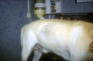
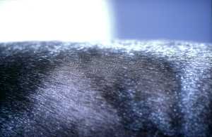
Bilateral non-inflammatory alopecias of the flanks are a relatively common reason for consultation in dogs.
Author: William Bordeau
Cabinet VetDerm,
1 avenue Foch 94700 MAISONS-ALFORT
They can result from dysendocrinias such as hypercorticism or hypothyroidism, but also from certain follicular dysplasias like canine recurrent flank alopecia (CRFA), also known as seasonal flank alopecia.
Follicular dysplasias constitute a rather heterogeneous group of genodermatoses, characterized by a structural anomaly of the hair follicle, leading to alopecia. A distinction is made between dysplasias that are linked to coat color (as is the case with black hair follicular dysplasia), and those that are independent of it. Likewise, a distinction is made between permanent follicular dysplasias and cyclic ones. Canine recurrent flank alopecia is a hereditary follicular dysplasia, which therefore falls within genodermatoses, and it is cyclic and not dependent on coat color. Given that it is a hereditary dermatosis, affected animals should not be bred.
Currently, almost nothing is known about its pathogenesis. At one point, it was speculated that hormones such as sex steroids, thyroid hormones, prolactin or corticosteroids were involved, but endocrine exploration of these animals has never revealed anything. However, it is thought that photoperiod may play a role. It is thus believed that a melatonin deficiency would be directly or indirectly responsible for this dermatosis.
This dermatosis affects a large number of breeds without sexual or racial predisposition, and without the castration or non-castration of the animal being a factor. It has been described in Airedales, Boxers, Bulldogs, Schnauzers, Poodles or the Bouvier des Flandres, among others. Affected animals are on average 4 years old, but the first lesions can appear between 8 months and 11 years.
This dermatosis classically manifests as hypotrichosis or alopecia well circumscribed with irregular margins giving an annular, polycyclic, or “geographical map” appearance. Hyperpigmentation is often observed. Hairs epilate easily in the affected areas and not elsewhere. There is no pruritus or pain. Squamosis and bacterial folliculitis may occur in the affected areas. This dermatosis is typically localized to the flanks, but can extend to the thorax and the dorsolumbar region. The involvement is usually bilateral, but sometimes one side may be more affected than the other. Exceptionally, the involvement may be unilateral. The extent of the lesions can vary over the years.
CRFA typically appears from November to March in the northern hemisphere, lasting for 3 to 8 months. In most cases, the hair regrows normally, however in some cases there may be a different texture or color. Sometimes, regrowth is incomplete after several episodes, and alopecia is thus almost permanent. This dermatosis is cyclic, but with variable cyclicity. Moreover, about 20% of animals experience only a single episode. Ultimately, this is a dermatosis whose name is inaccurate, especially because this dermatosis is not always recurrent and because it is not always localized to the flanks.
The differential diagnosis of symmetrical non-pruritic alopecias localized to the flanks is very important. Anagen and telogen effluvium, dilute color alopecia, black hair dysplasia, pattern alopecia, sebaceous adenitis, alopecia X, and all dysendocrine dermatoses must be considered. Even if they are generally not bilateral dermatoses, dermatophytosis and demodicosis should also be considered. The diagnosis of CRFA is based on anamnesis, historical data, clinical signs, exclusion of other dermatoses belonging to the differential diagnosis, and finally on skin biopsies. The lesions that can be observed are unfortunately not pathognomonic. It is only one element in favor of this dermatosis. Histopathological examination of biopsies shows diffuse follicular atrophy with dilated follicular infundibula filled with keratin, also present in secondary follicles and sebaceous glands, which gives a suggestive appearance known as “witch’s foot”. The sebaceous glands are generally normal in size or moderately hypertrophied.
The prognosis of this dermatosis is good, as it is not a dermatosis that can cause the animal’s death. The prejudice is only aesthetic. This varies with the importance of the extent of the lesions, the frequency of relapses, and the duration of each attack.
The extremely variable course of this dermatosis makes the evaluation of treatments all the more difficult. Thus, it should not be forgotten that even if nothing is done, in the majority of cases the hair regrows. Thus, it is difficult to say whether a molecule will decrease the frequency of attacks and their duration, since these can be very variable, spontaneously, in duration and in importance. Moreover, it should not be forgotten that it is a purely cosmetic problem. In any case, watchful waiting is a choice not to be neglected, and thus the use of molecules that can degrade the animal under the pretext of controlling this dermatosis will be avoided.

