

Brachycephalic dogs are enjoying growing popularity with the public, who are won over by their childlike features and endearing expressions. Behind this popularity, however, lies a heavy toll, which includes skin and ear disease. At the last ESVD congress in Lisbon, our colleague Elisa Maina gave a comprehensive review of their main dermatoses, setting out their mechanisms, clinical presentations and management.
These conditions are far from anecdotal: they affect between 10 and 30 % of individuals depending on the published series, and they result from a complex combination of genetic, conformational, environmental and microbiological factors.
Understanding brachycephaly: origins and a modern paradox
A revealing etymology
The term brachycephalic has its roots in Greek: brachy means short and cephale means head. This etymology alone sums up the morphological peculiarity of these dogs, whose skull is reduced in length and out of proportion to its width. This cranial disproportion is not merely an aesthetic detail: it shapes all the underlying anatomical structures, whether the upper airways, the hearing apparatus or the distribution of facial skin.
From functional to aesthetic selection
The history of these dogs goes back further than one might think. The first images of brachycephalic morphotypes date from the Assyrian period, between the seventh and ninth centuries BC. At that time, however, selection served a very different purpose from today’s: breeders sought maximum bite strength, useful for guarding or fighting.
The modern situation has been completely reversed. Today’s buyers generally no longer seek functional jaw strength, but the juvenile facial features typical of neoteny: round cheeks, large prominent eyes, a flattened muzzle. These features have been shown to trigger behavioural responses in humans similar to the maternal instinct, creating an almost irresistible attraction to these morphotypes. Aesthetic preferences are of course a personal choice that is not for a scientific setting to debate as such; it nevertheless remains essential to understand the psychological drivers of this attraction, because they directly explain why the phenomenon persists despite proven health risks.
The brachycephalic paradox
This popularity persists despite growing awareness of the health problems associated with this conformation. Behavioural studies of owners reveal a troubling phenomenon: many of them, even when buying the same breed again after a first dog affected by chronic disease, admit that they are fully aware of their animal’s suffering. This psychological mechanism, which could be described as cognitive dissonance applied to animal welfare, deserves careful reflection by the veterinary profession, whose educational role with future buyers remains decisive.
This conformation is indeed associated with reduced life expectancy, high morbidity and an objectively lower quality of life. Brachycephalic obstructive airway syndrome (BOAS) remains the best documented and most publicised condition. It should be remembered, however, that skin conditions are in no way less prevalent: in some breeds such as the English Bulldog, intertrigo is even more common than BOAS itself, with overall rates of dermatological disease exceeding 30 % of individuals.
Overview of dermatoses associated with brachycephalic conformation
The literature distinguishes two broad categories of dermatoses in these breeds. A key review thus provides a particularly complete and instructive inventory of all the skin conditions seen in these dogs, classifying them according to their causal link with craniofacial conformation.
The first category groups the conditions directly attributable to craniofacial conformation, precisely those that could in theory be reduced by less extreme breeding selection. The second category covers skin disorders that, although common in brachycephalic dogs, are not specifically linked to their morphology and are also seen in other breeds.
This distinction has considerable practical importance, because it guides prevention and genetic selection efforts: only the conditions in the first category can be reduced by a reasoned evolution of breed standards.
Dr Maina focused exclusively on the entities directly linked to brachycephaly: skin fold dermatitis, otitis externa, and primary secretory otitis media, also known as otitis media with effusion.
Skin fold dermatitis: anatomy of a friction disease
Terminology and definition
This condition goes by several names in the veterinary literature, notably skin fold dermatitis and skin fold pyoderma. It is fundamentally a surface pyoderma, meaning that it is characterised by excessive microbial proliferation, a cutaneous dysbiosis, without being a true infection in the strict sense.
The sites most often affected include the face, the vulva and the tail and peri-tail area, without excluding other locations where skin folds are present. Breed predisposition is clearly established in the English Bulldog, the French Bulldog and the Pug, breeds in which genetically selected body compactness produces excess skin that mechanically forms these deep folds.
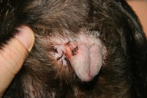
Vulvar intertrigo
Interestingly, the predisposition to excessive skin folds is not strictly limited to brachycephalic breeds in the narrow morphological sense. The Shar-Pei illustrates perfectly how excess skin, selected for its aesthetic appeal, systematically brings a train of complications: chronic secondary infection, persistent inflammation and a significant reduction in quality of life. This comparison with a breed that is not strictly brachycephalic underlines that the pathophysiological mechanism depends less on the shape of the skull itself than on the excess skin that often accompanies it, whether craniofacial in origin or spread more widely over the body.
Pathophysiology: when moisture meets friction
The pathogenic mechanism deserves a precise description, because it directly determines treatment choices. Saliva, tears, urine and sebum naturally tend to remain trapped at the bottom of these deep folds. This anatomical configuration, lacking effective ventilation, creates an almost permanently moist microenvironment that is particularly difficult for the owner to clean.
This chronic over-hydration combines with mechanical friction, generated by the constant rubbing of the two skin surfaces in contact. Together, moisture and friction progressively disrupt the stratum corneum. Once this skin barrier is damaged, skin permeability increases while local defence mechanisms are considerably weakened.
This degraded environment is an ideal breeding ground for bacterial and fungal proliferation. A mixed microbial overgrowth is thus seen, involving not only bacterial populations but also yeasts, notably of the genus Malassezia. This dysbiosis in turn sustains a local inflammatory response, which shows clinically as erythema and exudation. The microbial proliferation is often accompanied by a characteristic foul odour, and the resulting pruritus quickly becomes obvious to the attentive observer.
Without treatment, chronic lesions gradually develop: alopecia, hyperpigmentation, lichenification, erosions and ulcers complete this progressive picture. It should be stressed, however, that not all dogs with skin folds necessarily develop this dermatitis. This observation points to additional risk factors, among which concurrent allergies, obesity and certain hormonal imbalances appear to play a significant role in triggering clinical disease. Two dogs with morphologically similar folds can thus follow radically different clinical courses depending on whether these cofactors are present, which calls for a systematic assessment of the animal’s overall condition before concluding that this is simply an isolated mechanical problem.
A progressive spectrum of lesions
The clinical presentation of intertrigo follows a clearly identifiable continuum. In the early stages, only abnormal coat discolouration draws attention, caused by staining from the secretions accumulated in the fold. Over time, clearer signs appear: hyperpigmentation and lichenification reflect the chronicity of the inflammatory process.
In the most severe forms, skin maceration becomes obvious, with erosions, ulcers and crusts, sometimes together with extensive alopecia. This gradation justifies a systematic and careful clinical examination of all the folds, including those not immediately visible on superficial inspection. A superficial examination limited to the most accessible areas risks missing deeper and more advanced lesions hidden at the bottom of particularly tight folds.
Ocular complications of facial folds
Skin folds on the face are not just a dermatological problem: they also expose the animal to significant ophthalmological complications. When excess folds are directed towards the periocular region, the hairs emerging from them can come into direct contact with the corneal surface. This chronic contact first causes traumatic keratitis, which can progress to frank corneal ulceration if the mechanical factor persists. This association between excessive facial folds and corneal disease has been formally reported in brachycephalic breeds, reinforcing the need for a systematic ophthalmological examination in any patient with facial skin fold dermatitis.
The special case of the screw tail
The tail region deserves particular attention, especially in the English Bulldog, in which the conformation of the ingrown or screw tail creates an environment conducive to the accumulation of organic matter. Unlike the facial fold, which retains some flexibility, the tail is constrained here by a rigid bony structure that considerably limits its mobility. This relative immobility encourages hairs to become buried and increases local friction.
Clinically, secondary infection causes pruritus that leads the animal to rub, bite or lick the affected area, producing further secondary lesions. Some cases reach remarkable severity, with yellowish, dry, probably purulent material hidden at the bottom of the fold and sometimes invisible without close examination under suitable magnification. This discretion explains why many owners do not realise how serious the situation is until severe ulcers, with deep infection, become obvious.
This tail anatomy is often accompanied by further complications. When the tail physically obstructs the anal region, faeces can become trapped, causing first perianal dermatitis, then potentially anal sac disease, or even a risk of perianal fistulae. This cluster of complications is seen mainly in Bulldog-type breeds, but it has also been reported in the Pug and the Boston Terrier.
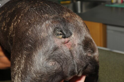
Severe tail fold intertrigo
Other sites: lips, neck and vulva
Although less characteristic than in the French Bulldog, problematic skin folds can also occur in other breeds, such as the Cocker Spaniel, in which lip or neck fold dermatitis can develop through an identical pathophysiological mechanism.
The vulvar region is a particularly interesting site because the animal can reach it to lick. The resulting chronic licking progressively worsens the initial lesions. Early stages are limited to abnormal coat discolouration, with mild hypotrichosis and sometimes a small focal ulcer. Chronic progression leads to more numerous and more diffuse lesions.
This vulvar location carries notable systemic and functional complications. Chronic irritation from the vulvar fold promotes ascending urinary tract infections. It should be noted that this association is not linked exclusively to brachycephalic conformation as such, but rather to the persistence of a juvenile vulvar conformation. One therapeutic point deserves emphasis: surgical resection of the vulvar fold brings a proven improvement in both recurrent urinary tract infections and the vulvar dermatitis itself, arguing for a surgical approach in refractory cases.
Diagnostic approach
The diagnosis of intertrigo rests first and foremost on clinical examination. The strictly intra-fold location of the lesions is in itself a strong diagnostic element, making this an essentially clinical diagnosis. Systematic cytology nevertheless confirms the presence of bacteria or yeasts and guides the choice of treatment according to the predominant microbial population.
The clinician must, however, remain alert to certain differential diagnoses or concurrent conditions. When intertrigo affects the face, concurrent atopic dermatitis must always be sought, because it is a major predisposing factor for relapses of skin fold dermatitis. Atopic dermatitis rarely shows only in the folds: looking for other typical sites, such as ventral dermatitis, otitis or conjunctivitis, is essential to establish this concurrent diagnosis.
Skin biopsies and histopathology are rarely indicated in this condition. They do, however, have a place in certain particular situations, notably when conditions such as mucocutaneous pyoderma or mucocutaneous lupus erythematosus, in subtle forms, can be confused with severe skin fold dermatitis, especially on the vulva. In these ambiguous circumstances, histology can resolve the diagnostic doubt.
Treatment principles: a stepwise, reasoned approach
Treatment of intertrigo should logically follow the stages of its pathophysiology. Three successive lines of treatment should be considered: control of microbial proliferation and infection, control of inflammation and, finally, management of residual moisture and friction.
For microbial control, the choice of treatment should be guided by the clinical presentation and cytology results. Mild forms, with moderate bacterial proliferation, generally respond to simple cleaning with antimicrobial wipes, sprays or suitable shampoos. More severe clinical forms, with a large population of cocci on cytology, justify applying a mupirocin ointment twice daily after prior cleaning. In severe cases where the inflammation is mixed, silver sulfadiazine is an appropriate alternative. Finally, deeply ulcerated tail lesions with frank, deep infection require systemic antibiotic therapy, always guided by culture and susceptibility testing to avoid any inappropriate empirical prescription.
Control of inflammation, by contrast, is less well codified in the veterinary literature, owing to a lack of sufficient studies in both human and veterinary medicine. It is nevertheless accepted that topical corticosteroids, and systemic corticosteroids in the most resistant forms, are potentially effective in these chronic conditions in which the inflammatory component plays a decisive role. A proactive, preventive strategy can help reduce the frequency of relapses, bearing in mind that this condition is chronic in the absence of definitive surgery. Managing concurrent conditions at the same time, particularly atopy when present, is essential for lasting control of the disease.
The third pillar of treatment concerns control of moisture and friction, achieved with drying agents and barrier creams, of which zinc oxide is the most representative example. The application technique must follow a strict sequence: thorough cleaning of the fold, which is usually very soiled, must always come before applying the barrier cream or drying agent. Using chlorhexidine wipes followed by a zinc oxide paste illustrates a practice commonly recommended to owners, to be carried out with protective gloves, to avoid any prolonged direct contact with the secretions and topical products used.
In some cases, corrective surgery of excessive folds is a treatment option not to be overlooked, particularly when conservative medical measures fail to control the condition in the long term. This procedure, although invasive, brings a substantial improvement in the animal’s quality of life by permanently removing the anatomical substrate responsible for the disease.
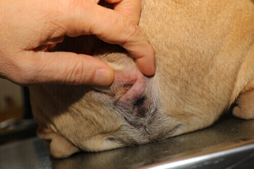
Corrective surgery is sometimes needed, but not always feasible
Otitis externa in brachycephalic dogs: a multifactorial disease
General pathophysiological reminder
Otitis externa is defined as an inflammatory condition of the external ear canal and the pinna. Its pathogenesis is classic: predisposing factors, primary causes and perpetuating factors. In brachycephalic breeds this conceptual framework remains valid, but each component is considerably amplified by the anatomical features of this conformation. In other words, in brachycephalic dogs it is not so much the nature of the factors involved that differs, but their intensity and how they interact, which makes managing this condition particularly complex in this population.
Anatomical features that worsen the prognosis
A normal ear canal should allow the tympanic membrane to be seen easily on otoscopic examination. In brachycephalic dogs, this is often impossible. One study reports that in a sample of 150 ears examined, the tympanic membrane was visible in only five, that is, it could not be seen in 145 ears out of 150. An additional CT assessment measured the diameter of the ear canal objectively, showing it to be half that of dogs with mesocephalic conformation. The ear canal was thus partially or completely occluded in 82 % of the cases studied. It should be noted that this particular study did not establish a direct statistical correlation between the degree of stenosis and the prevalence of otitis externa; other studies nevertheless confirm an overall higher prevalence of this condition in brachycephalic breeds than in other skull types.
The anatomical abnormalities are not limited to the external ear canal. The tympanic bulla itself shows notable morphological features in these breeds: it is smaller, thicker and positioned more rostrally. These structural changes can impair the function of the Eustachian tube, predisposing to a higher frequency of otitis media which, paradoxically, often escapes detection on standard otoscopy, precisely because stenosis of the external ear canal prevents any direct view.
The combination of these anatomical features leads to reduced ventilation of the ear canal. This poor ventilation mechanically promotes retention of cerumen, creating a microenvironment favourable to microbial proliferation. When concurrent allergic disease, a source of chronic inflammation, is added to this already unfavourable terrain, it is easy to see why these animals develop tissue hyperplasia and fibrosis more readily than other conformations. It should also be remembered that middle ear effusion, often silent because it cannot be seen, helps delay the diagnosis of the otitis externa itself, leading to more severe clinical pictures at the time of presentation.
Otitis media in brachycephalic dogs
Defining otitis media and its breed-specific features
Otitis media is characterised by a pathological change in the tympanic bulla, identifiable on imaging. This change may involve the lumen of the cavity, with intracavitary effusion, or the bone itself, with bone proliferation or lysis, and both pictures may coexist.
A fundamental distinction separates the pathophysiology of otitis media in dolichocephalic and mesocephalic breeds from that seen in brachycephalic dogs. In the former, involvement is classically secondary to pre-existing otitis externa, with infection spreading from outside inwards and progressive damage to the tympanic membrane: purulent material accumulated in the external canal eventually breaches the integrity of the eardrum, allowing the infection to extend into the tympanic bulla itself. Reported rates of progression vary with the type of initial otitis externa: 20 % for erythemato-ceruminous otitis externa, 50 % for suppurative otitis and up to 70 % for proliferative otitis.
In brachycephalic dogs, by contrast, this classic ascending mechanism does not appear to predominate. Most of these animals show no significant inflammation of the external ear canal, despite a confirmed effusion in the tympanic bulla. This clinical dissociation strongly suggests a primary pathophysiological mechanism, independent of any prior external disease and directly linked to the anatomical features of this skull type.
A shifting but revealing terminology
The literature on this entity uses variable terminology, reflecting the gradual evolution of pathophysiological understanding. First described in the Cavalier King Charles Spaniel as primary secretory otitis media, the condition was later reported in other brachycephalic breeds as otitis media with effusion, a term probably borrowed from human medicine, where this paediatric entity is often accompanied by a triggering bacterial component, or at least a well-identified bacterial trigger in children.
In dogs, however, clinical observations show that inflammation and infection are not always present, which has led some authors to prefer the more neutral term middle ear effusion, which is more faithful to the canine pathophysiological reality and avoids any unwarranted infectious connotation.
Different pathophysiological mechanisms by breed
In the Cavalier King Charles Spaniel, the particular pharyngeal conformation is associated with Eustachian tube dysfunction, which impedes the physiological drainage of mucus produced within the tympanic bulla. This dysfunction creates negative pressure in the middle ear, which is thought to exert a suction effect on cellular secretions, promoting increased production of viscous mucus.
In the other brachycephalic breeds concerned, namely the Boxer, the Boston Terrier and the French Bulldog, the pathophysiology appears multifactorial. A narrow Eustachian tube, particularly well-developed head and neck muscles that may mechanically interfere with tubal opening, temporomandibular joint dysplasia limiting how wide the mouth opens during chewing, an elongated soft palate, and abnormal size and position of the tympanic bulla are all contributing factors. Some studies also report altered goblet cell production, with effects on breathing patterns that may further increase negative pressure in the middle ear. It is therefore a combination of anatomical and functional factors that explains the accumulation of secretions in the middle ear in these breeds.
A clinical picture that varies by breed
The available data on the clinical expression of this condition are uneven across breeds, which calls for some caution in interpretation. The Cavalier King Charles Spaniel has the richest documentation, whereas there is only one dedicated study in the Boxer, a single publication on just two cases in the Boston Terrier, and the French Bulldog is only beginning to be investigated more systematically.
One fundamental clinical point deserves emphasis: middle ear effusion can remain entirely asymptomatic. This incidental finding, made during head imaging performed for another reason, concerns more than 50 % of French Bulldogs examined. By contrast, the Boston Terrier has always been symptomatic in the reported cases.
Neck and head pain is a particularly early and significant clinical sign in the Cavalier King Charles Spaniel. The Boxer, for its part, shows pain on opening the mouth, while head shaking is seen more or less consistently in all the breeds concerned, with the notable exception of the Boston Terrier. Hearing loss and deafness are also among the reported signs, and are particularly important in the Cavalier King Charles Spaniel, in which they appear early in the clinical course.
Vestibular signs and facial paralysis complete this clinical picture. These neurological signs result from compression by mucus or purulent material accumulated in the bulla, from the direct action of toxins, or from inflammatory lesions of the cranial nerves crossing the tympanic region or the adjacent vestibular structures. Vestibular signs are particularly severe in the Boston Terrier, whereas they vary in the other breeds. Facial paralysis has also been reported in several breeds, although its presentation remains uncertain in the French Bulldog for lack of a sufficiently detailed clinical description in the available literature. Chewing difficulties have also been described in the Boxer, while concurrent otitis externa affects only a minority of cases, such as a single case out of seven reported in this breed.
A structured diagnostic approach
Clinical suspicion of otitis media rests initially on the history and the animal’s clinical signs. Video-otoscopy is the essential next step, allowing assessment of the integrity of the tympanic membrane, a search for foreign bodies, evaluation of any bulging, and detection of signs of previous damage such as membrane opacities.
Breed-specific clues have been reported regarding the appearance of the tympanic membrane. In the Cavalier King Charles Spaniel with middle ear effusion, a characteristic bulging of the pars flaccida has been described. This differs from the Boxer, in which bulging preferentially affects the pars tensa according to the data reported by Patterson in the cases he described. The Boston Terrier, for its part, shows no notable bulging of the membrane. Data on the French Bulldog remain largely incomplete, as the tympanic membrane usually cannot be seen in this breed because of severe stenosis of the ear canal.
Imaging usefully complements otoscopy. CT has excellent sensitivity for detecting effusion, assessing the morphology of the bulla, detecting bone changes and judging the chronicity of the lesions. MRI, on the other hand, gives a better view of soft tissues, the inner ear and nerve structures, and is particularly relevant when there are concurrent neurological signs, notably in the Cavalier King Charles Spaniel when associated neurological disease is suspected.
Imaging nevertheless has certain limitations that must be borne in mind. The high rate of incidental findings in the French Bulldog, more than 50 % of cases, unilateral or bilateral, calls for cautious interpretation of the results. It therefore seems sensible to use imaging only when clinical suspicion of otitis media rests on sufficiently convincing historical and clinical evidence, thus avoiding over-interpretation of incidental findings of no proven clinical relevance.
Myringotomy, also called tympanic paracentesis, is the next diagnostic and therapeutic step. This technique confirms the presence of effusion while allowing a sample to be taken for cytology and bacteriology. The incision can be made with a catheter or a small blade, after which a catheter is introduced to collect the sample for cytology and for culture and susceptibility testing. Subsequent flushing must be thorough and persistent, as the mucus is sometimes particularly dense and resists simple rinsing; it is not unusual for the clinician to have to repeat the flushing several times before the accumulated secretions are satisfactorily cleared.
The preferred site for this myringotomy remains the caudoventral pars tensa, provided the tympanic membrane is actually visible, a condition rarely met in the French Bulldog, in which access must be adapted to individual anatomical constraints.
One technical point deserves particular attention: positioning of the animal during this procedure. A recent study compared contamination rates according to the approach used: the traditional approach through the lateral ear canal carries a contamination rate of 67 %, whereas an alternative position, in lateral recumbency with vertical access from beneath a specially designed table, reduces this rate to just 5.5 %. This considerable difference strongly supports adopting this alternative technique in routine practice, to ensure reliable bacteriology results.
Gross appearance of the effusion by breed
Comparison of middle ear effusion reveals notable differences between breeds. In the Cavalier King Charles Spaniel, the Boxer and the Boston Terrier, the effusion usually takes the form of a particularly viscous and tenacious mucus plug, varying in colour from yellowish through greyish to whitish.
The French Bulldog, by contrast, shows much greater gross variability: the effusion may be mucoid, serous and honey-coloured, but also brownish or frankly bloody. This diversity is probably explained by the more frequent presence of inflammatory cells together with bacteria in this breed, with positive cultures in about 50 % of reported cases. This more marked inflammatory and infectious component could explain the generally more fluid consistency of the effusion when it is purulent. Conversely, the effusion is mostly acellular in the Cavalier King Charles Spaniel and the Boxer, in which bacteria are almost always absent from the samples analysed.
Treatment options and management of recurrence
Recurrence of the effusion, particularly common in the Cavalier King Charles Spaniel, can be managed by repeat myringotomy. In most recurrent cases, however, placing a tympanostomy tube, also called a grommet, is a more lasting solution. This very small device, placed in the centre of the tympanic membrane, acts as a true doorway for secretions: it allows continuous drainage of the fluid produced, preventing its gradual accumulation in the tympanic bulla.
After the initial flush, medical treatment relies on several drug classes. Anti-inflammatories, notably prednisolone, can be given topically, orally or by both routes. Antibiotic therapy, on the other hand, should only be started when cytology and bacteriology are positive, as the effusion is sterile in many cases, particularly in the Cavalier King Charles Spaniel and the Boxer. Finally, N-acetylcysteine, a recognised mucolytic, is of definite therapeutic interest because it breaks the glycoprotein bonds of mucus, making it more fluid and easier to drain. It can be given topically or systemically depending on the clinical severity.
Prognosis by breed
The prognosis of this condition varies considerably by breed. In the Cavalier King Charles Spaniel, the chronic, recurrent course significantly impairs quality of life and requires lifelong management. The Boxer, by contrast, generally has a favourable prognosis, with recurrence rare in this breed. The Boston Terrier shows a variable course: mild forms may improve spontaneously, whereas severe forms may leave permanent neurological sequelae. Finally, current knowledge of the French Bulldog remains insufficient: the often incidental nature of the finding raises questions about the true clinical relevance of this entity in this breed, a question that still lacks a definitive answer and deserves further investigation.
Conclusion
Far from being a mere aesthetic trait sought by owners, brachycephalic conformation brings a deep anatomical predisposition to a range of dermatological and otological diseases whose severity and chronicity call for constant clinical vigilance. Skin fold dermatitis, otitis externa and primary secretory otitis media, although their clinical expression varies between breeds, share a common origin: the structural anatomical change imposed by the extreme morphological selection of these dogs.
Early recognition of these changes remains decisive in putting in place appropriate treatment and proactive management that limits the development of irreversible chronic lesions. Veterinarians should not hesitate to propose surgery, particularly for major skin folds or recurrent middle ear effusions, when conservative medical measures fail to control the signs in the long term.
Our collective responsibility is also to inform breeders and future owners rigorously, so as at least to encourage the selection of less extreme types within the brachycephalic breeds themselves. Brachycephaly cannot be reduced to a simple aesthetic attribute: it carries a tangible health cost, affecting many of the animal’s physiological systems. Further work, particularly on the French Bulldog, for which data are still fragmentary, would refine our understanding of the true clinical relevance of certain incidental imaging findings and help harmonise diagnostic and treatment protocols across the breeds concerned.
Maina E. Dermatological issues in brachycephalic dogs. Lisboa, ESVD Meeting 2025
