

Claw disorders remain uncommon in dogs and cats: Dr Zanna puts them at around 1 to 3 % of veterinary consultations, a proportion that varies with the population examined. They are often under-reported or overlooked, because they seem minor until they are recognised as the signal of a wider dermatosis or disease.
The claw is not an isolated appendage: its matrix is richly vascularised, and a deformity, a separation or a softening may be the first visible sign of a neoplasm, an immune disorder or a systemic infectious disease. At the ECVD-ESVD congress in Bilbao, our colleague Giordana Zanna (Istituto Veterinario di Novara, Italy) discussed claw diseases in dogs and cats, both their aetiology and their management.
1 The vocabulary of claw disorders
A set of specific terms is used to describe claw disorders in dogs and cats, largely borrowed from human medical terminology. Dr Zanna notes that she found differences between human and veterinary usage for some of these terms, which means the intended meaning should be stated each time.
Paronychia refers to inflammation of the tissues surrounding the claw, whereas onychitis refers to inflammation of the claw itself. This inflammation can lead to onychomadesis, that is, shedding of the claw. A claw that does not fall off may soften: this is onychomalacia, a softening that can be felt simply by touch.
Onychoschizia refers to splitting, or lamination, of the claw. This splitting occurs in the horizontal plane, at the free edge, and Dr Zanna explains that she understood it this way after comparing human and veterinary definitions. Onychorrhexis, by contrast, is recognised by longitudinal striation, associated with brittleness and breakage of the claw.
In onychocryptosis, the claw overgrows, curves inwards and eventually becomes ingrown into the adjacent digital pad. The pain is constant. Onychogryphosis, the most familiar to practitioners, refers to an abnormally curved, deformed and hypertrophied claw. To these terms is added onychodystrophy, which refers to abnormal claw formation. Onycholysis, classically the separation of the claw plate from its bed, will come up in several contexts.
Chromonychia, a term that Dr Zanna says she came across in an Italian textbook and that was adopted from human medicine, refers to abnormal colouring of the claw.
2 The approach: never isolate the claw from the animal
A claw disease is approached exactly like any other dermatological condition. Dr Zanna places two symmetrical rules at the heart of her approach: do not focus on the claw alone, and do not ignore the rest of the animal. In other words, the full history, the general dermatological examination and routine ancillary tests come before the examination of the claws, not after it.
Isolated paronychia is rarely a diagnosis in itself; it points towards a hypersensitivity, demodicosis, an immune disorder or a systemic condition, which only examination of the rest of the skin can identify. Combining a thorough history, the clinical examination and diagnostic techniques (cytology, biopsies, imaging) leads to a more accurate diagnosis, and it is this diagnosis that allows treatment to be directed at the underlying cause rather than at its manifestation in the claw.
Whether one claw or several are affected guides the reasoning from the outset. Conditions affecting one or a few claws asymmetrically typically reflect a localised problem: trauma, bacterial or fungal infection, neoplasia. Conversely, when several claws, or all the claws on different feet, are affected, the causes are often systemic: autoimmune diseases, immune-mediated conditions, nutritional deficiencies, congenital abnormalities, or infections resulting from a systemic disease.
3 Anatomy of the claw and the value of imaging
Before discussing tumours, one must know the normal in order to read the abnormal on a radiograph. Dr Zanna draws these landmarks from her regular exchanges with radiologists and other specialists.
In a normal claw, the ungual crest is a cone-shaped bony shell carried by the third phalanx. The ungual process, for its part, is a pointed bony projection. The claw itself is anchored on this assembly. Three structures, then: the distal phalanx, the crest that caps it and the process that extends it. The claw attaches to them.
These landmarks become diagnostic criteria in subungual keratoacanthoma, in which the radiologist regards three features as the hallmark of the lesion. First, the body of the third phalanx is spared: osteolysis is concentrated on its two distal projections. Second, the ungual crest undergoes expansile osteolysis: it is eroded and deformed outwards. Third, the ungual process undergoes complete osteolysis, accompanied by widening of the claw. Two recent radiographic case series find this trio (severe resorption of the process in 12 of 12 dogs and 7 of 7, expansion of the crest in 10 of 12 and 4 of 7, widened claw in 11 of 12 and 7 of 7), but they also report lysis of the distal articular surface of the second phalanx in 6 of 12 dogs in one series and 2 of 7 in the other: the distal phalanx is therefore not “normal”, only its body is. Taken together, these three signs allow the radiologist to raise a strong suspicion of subungual keratoacanthoma, which in practice guides surgery and analysis even before the histological result.
4 A single claw affected
When a single claw is affected, two causes predominate: traumatic injury and tumour.
4.1 Traumatic injury
The trauma occurs in ordinary circumstances (after a run, a day’s hunting) and its picture is unmistakable: claw abnormality, marked pain, bleeding. Over time, the same claw may become secondarily infected, and the secondary bacterial infection may spread to the interdigital area. Exudate is then seen either on the claw alone or also on the interdigital skin.
The fractured portion must be removed, while preserving the area of living tissue, the matrix, which is richly vascularised and nourishes the claw: it is the matrix that will allow regrowth. Antiseptic soaks are applied; they can be irritating. Analgesics and a protective dressing are sometimes needed. Finally, in the event of secondary bacterial infection, systemic antibiotic therapy is not needed, as topical treatment is normally sufficient. This position, put forward in the lecture without a cited reference, reflects Dr Zanna’s clinical experience.
4.2 Subungual keratoacanthoma
Claw tumours are numerous; Dr Zanna describes three. The first is subungual keratoacanthoma, whose radiological signs were described above. The histological counterpart of these images confirms the lesion.
4.3 Subungual squamous cell carcinoma
The diagnosis of subungual squamous cell carcinoma is pathological, mainly histopathological. On examination, the cells appear polygonal, with anisokaryosis and anisocytosis. The tumour occurs mainly in certain breeds, shown on screen but not named in the transcript, and it has also been described in cats.
4.4 Subungual malignant melanoma
Subungual malignant melanoma can affect claws that become discoloured. Examination reveals characteristic cells on a pigmented background. Here too, the description mainly concerns certain breeds, not named in the transcript. Dr Zanna reports a recent case in which the claw had been completely destroyed by the tumour.
4.5 Principles of management
Cooperation with the radiologist, presented as essential, makes it possible to understand what is happening in the digit and the claw, beyond what inspection shows.
In all these cases, the best approach is digit amputation. Submitting the claw alone, the detached claw, is of no use: one must be able to determine what is happening in the bone itself and whether the tumour has metastasised elsewhere.
5 One or two claws affected: onychomycosis
When one or two claws are affected, onychomycosis is among the best differential diagnoses. The picture combines frank onychomalacia (the claws are completely soft) and onycholysis. Involvement normally remains limited to one or two claws.
Dr Zanna illustrates this with a case in which digital involvement preceded claw involvement: a dog infected with Trichophyton mentagrophytes, in which the infection first affected the digits before spreading to the claw itself, again with onychomalacia. This sequence is a reminder that the claw can be affected by contiguity from the digital skin, and that examining the digits is part of examining the claw.
Diagnosis relies on fungal culture. The sample is taken from the proximal part of the claw, that is, the matrix side, which offers the best diagnostic accuracy. The claws are broken into fragments, then placed on a routine dermatophyte culture medium. A claw biopsy is not mandatory when culture is sufficient.
Antifungal treatment relies on the available drugs, used alone or in combination. Treatment must be continued for several months before resolution is achieved at the claw. Dr Zanna indicates that complete regrowth sometimes takes six to seven months. This duration must be explained to the owner from the outset, because it determines compliance.
6 Multiple claws in an otherwise healthy animal: symmetrical lupoid onychitis
When almost all the claws are affected in an otherwise healthy individual, the most important diagnosis is symmetrical lupoid onychodystrophy, the most common of the diseases that cause symmetrical onychomadesis and onychodystrophy in dogs.
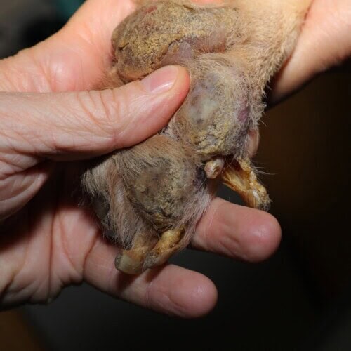
Severe symmetrical lupoid onychodystrophy
6.1 A question of name, which is a question of mechanism
Dr Zanna challenges the established name, for a pathophysiological reason. This is not a disease of primary claw formation. The claw changes are normally secondary to inflammation of the claw bed, which causes the claws to separate; only afterwards, secondarily, does regrowth become abnormal, producing deformed and brittle claws. Since inflammation precedes and causes the dystrophy, it would be preferable to speak of symmetrical lupoid onychitis rather than symmetrical lupoid onychodystrophy.
6.2 Clinical presentation
In the same dog, several feet are affected, and the lesions combine: onycholysis, onychomalacia, onychodystrophy and paronychia coexist.
6.3 Predispositions and aetiological hypotheses
The disease has mainly been described in certain breeds, in which its prevalence is higher. A genetic predisposition is likely.
Class II haplotypes of the dog leukocyte antigen, involved in the immune response, have been studied as candidate genes.
A link with adverse food reaction has also been considered, based on a prospective study of 24 dogs with exclusive claw involvement: four achieved partial or complete resolution on an elimination diet; two of them underwent a provocation test and relapsed, while the other two were not challenged. Dr Zanna considers this proportion very small and the link unproven; the study’s authors, however, concluded that the elimination diet is a diagnostic test worth performing, and a later systematic review lists symmetrical lupoid onychitis among the reported non-cutaneous manifestations of adverse food reaction.
A third hypothesis concerned raised levels of antithyroid antibodies, which the authors suggest might also affect the claw matrix. Here again, Dr Zanna considers the evidence for this link insufficient. None of these three leads establishes the aetiology.
6.4 Diagnostic approach
Symmetrical lupoid onychitis presents as a distinct disease in an otherwise healthy dog. Haematology and biochemistry are performed as a general baseline, to assess overall health, before starting treatment, not to make the diagnosis, but to make the subsequent treatment safe.
That leaves biopsy. The technique discussed here was introduced many years ago, and it applies not only to symmetrical lupoid onychitis but to many other claw disorders. It is simple. An 8-millimetre biopsy punch is positioned horizontally, meaning that the cutting edge is perpendicular to the sagittal and transverse planes. Oriented in this way, it passes through both the claw matrix and the underlying third phalanx. The technique therefore samples both structures in a single cut: the claw bed and its bony support are read together, on the same slide, with the claw plate opposite.
6.5 Histology
The characteristic lesion is a lichenoid band of lymphocytic infiltrate. Vacuolation of basal cells has also been described, but this finding is very common in many other inflammatory diseases and is therefore not specific to symmetrical lupoid onychitis.
6.6 Treatment goal and treatments
The first step of treatment is a conversation with the owner. The goal to set is strong, comfortable claws, not cosmetically perfect claws: the latter result is impossible to achieve. Setting this goal from the outset avoids the dissatisfaction of an owner who would judge failure by appearance.
Treatment combines several modalities, shown on screen, with the dose of the first depending on the dog’s weight. Other treatments have been used in combination or alone. No monotherapy and no combination appears more effective than the others. The choice therefore rests on clinical judgement and on the animal’s tolerance.
The condition is managed over time rather than cured.
7 Multiple claws with paronychia
When multiple claws are affected with paronychia in the foreground, two signs point the way: waxy debris and a brownish waxy discolouration. This abnormal colouring is chromonychia. The presence of associated skin lesions strengthens the suspicion.
7.1 Malassezia overgrowth in dogs
Faced with this picture, Malassezia infection or overgrowth must be considered. Dermoscopy clearly shows the brownish waxy material around the claw fold, as well as the brownish discolouration of the claw itself.
Diagnosis relies on the basic tests used to demonstrate Malassezia. The material can be scraped off and examined: the small, round bodies characteristic of the yeast are then seen.
Management of Malassezia paronychia, or of associated chromonychia, only succeeds if the underlying cause of the overgrowth is treated. These dogs normally also have other hypersensitivity disorders. The primary disease must therefore be treated, rather than focusing solely on the yeast overgrowth. If Malassezia is nevertheless to be treated, treatments based on concentrated antiseptics are preferred as first line.
Dermatological problems that first affect the interdigital skin, those linked to Malassezia spp. infection and associated with hypersensitivity disorders, can change the colour of the claws and extend their effects to the claw folds, with accumulation of brown, waxy keratinous debris.
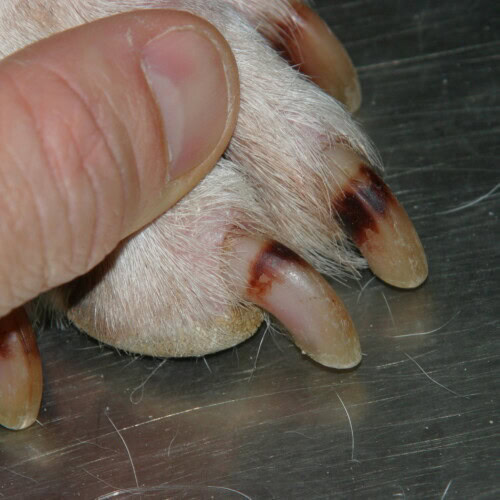
Change in fungal discolouration after starting ketoconazole
7.2 Malassezia overgrowth in cats
This Malassezia-associated paronychia or chromonychia is also described clinically in cats, mainly in the Sphynx and the Devon Rex. Dr Zanna notes that Sphynx breeders are fully familiar with this type of lesion.
In these cats, the claw fold itself, comparable in principle to what is seen in the Shar-Pei, may predispose by maintaining chronic moisture and friction. But Dr Zanna holds to the general rule: most of these cats also have underlying hypersensitivity disorders, as the most typical cause. This Malassezia overgrowth has also been described in cats with visceral paraneoplastic syndromes.
Diagnosis therefore relies first on identifying the underlying disease, then on treating the overgrowth itself. Finally, the abundance and diversity of Malassezia species depend on the site: Malassezia slooffiae has mainly been described in the claw folds.
7.3 Bacterial paronychia
Any underlying skin condition can induce bacterial paronychia. Cytology then reveals the cells expected in bacterial inflammation. In cats, bacterial paronychia is less common and is normally associated with other underlying skin diseases. The same logic applies as for the yeast, then: paronychia is the symptom, not the diagnosis.
7.4 Demodicosis
Some skin conditions mainly affect the folded parts of the digits and can secondarily reach the claw fold: demodicosis is an example. Dr Zanna reports the case of a dog with demodicosis in which microscopic examination confirmed the diagnosis and whose claws were long because of inflammation of the claw matrix.
8 Multiple claws with onychogryphosis
When onychogryphosis dominates the picture, two differential diagnoses stand out: leishmaniosis and ischaemic dermatopathy.
8.1 Leishmaniosis
Leishmaniosis can present with onychogryphosis, but in many cases other distinctive signs accompany the claw involvement. The lesions observed combine dystrophy and onychogryphosis, up to cases in which all the claws are affected and curved.
Why do these dogs develop onychogryphosis? An article published many years ago sought the answer by focusing on the claws of dogs with leishmaniosis. Forty dogs were divided into two groups: 16 of 40 had generalised onychogryphosis, 24 of 40 did not. All the claws were sampled, and PCR and histochemistry were performed on these samples.
The claw must be decalcified, a technique that limits antigen expression; the PCR results are therefore difficult to interpret. The authors nevertheless performed it: Leishmania was detected by PCR in 3 of the 16 dogs with onychogryphosis (denominator missing from the transcript, restored from the original publication), and in 2 of 24 dogs in the group without onychogryphosis. Histopathology, performed in parallel, revealed a pattern of lichenoid onychitis, characteristic of an inflammatory condition.
Onychogryphosis is not accompanied by substantial local parasitism and probably results from the systemic infection, or from the chronic immune activation it sustains. A smaller series, 7 dogs with onychogryphosis and 9 without, studied by immunohistochemistry, has since detected amastigotes in the dermis adjacent to the matrix, more often in the dogs with onychogryphosis; its size and its publication outlet carry less weight than the original study, but the question of local parasitism is not closed. The deformed claw reflects a systemic state, not colonisation of the claw, and looking for the parasite in the claw is unlikely to succeed.
8.2 Ischaemic dermatopathy
Onychogryphosis can also occur as part of ischaemic dermatopathy. The term is very broad and covers many conditions sharing the same clinical and histopathological findings.
A vascular change reduces blood flow; the reduced flow causes tissue hypoxia; the hypoxia compromises the claw matrix and consequently causes claw dystrophy. The claws then become soft, lack support, weaken and flake.
According to Dr Zanna, the treatments that can be used are those already mentioned for symmetrical lupoid onychitis. But the diagnostic approach differs: faced with ischaemic dermatopathy, an underlying disorder must be sought, and she stresses a point that has only recently become widely recognised: dogs with leishmaniosis can develop ischaemic dermatopathy. Leishmaniosis is therefore not only a differential diagnosis of onychogryphosis; it is sometimes its cause, by way of ischaemic dermatopathy.
9 Rapid claw growth
Rapid claw growth can occur in several settings, for which Dr Zanna gives four examples linked by the same reasoning: accelerated turnover, or inflammation of the matrix.
The first concerns the hyperthyroid cat: a colleague specialising in internal medicine observed these cases, which Dr Zanna explains by increased metabolic turnover. The second relates to behaviour: animals whose scratching activity is reduced, especially older animals, can also have long claws. The third is paronychia: in a dog with paronychia, any underlying skin condition can induce bacterial paronychia and, for this reason, cause rapid claw growth. The fourth, already mentioned, is demodicosis, in which the claws were long because of matrix inflammation.
The practical value of this list is to remind us that a claw growing too fast is a sign to investigate, not simply a reason for a trim.
10 Onychomadesis and rare conditions
Multiple claws can also be affected as part of other, rarer underlying conditions, in which onychomadesis is often the presenting sign.
Genodermatoses are among them. Junctional epidermolysis bullosa, covered elsewhere at the congress, can be accompanied by onychomalacia; the disease also affects other areas, notably the mouth and ears. The mechanism is structural: mutation of a structural protein disrupts the integrity of the claw matrix and claw bed. Dr Zanna cites a figure of 5 % in this connection, but what it refers to cannot be made out from the transcript.
Immune-mediated diseases can also affect several claws, with a low degree of involvement. Pemphigus foliaceus is an example, with serous paronychia and onychomadesis. In cats, the picture is even clearer.
Finally, when onychomadesis predominates, certain rare diseases must be considered: subepidermal blistering diseases, genodermatoses, or pemphigus in cats.
11 Claw involvement in cats
Cats are mainly affected secondarily by claw diseases. Dr Zanna discusses two of them, noting that the same digit may carry lesions that are difficult to interpret: bloody and crusted material inside the claw, onychomadesis, paronychia affecting several claws.
11.1 Feline lung-digit metastatic syndrome
According to Dr Zanna, cats have a rich blood supply to the digits, which she links to thermoregulation.
The primary tumour may be a bronchial or bronchoalveolar adenocarcinoma, with obvious angioinvasive behaviour. Digital involvement is secondary to haematogenous spread, through arterial embolisation of cells from the primary tumour, and the rich digital blood supply explains this location. The weight-bearing digits are normally affected.
Diagnosis requires first demonstrating the lung tumour, then confirming it by cytology, sometimes also by histology; ideally, the same cells should be found in the lung and in the digits to attribute both lesions to the same disease. Respiratory distress in these cats normally appears after the first dermatological signs affecting the claws. The cat is therefore presented for digital pain, and it is this claw disease that is subsequently traced back to the lung tumour.
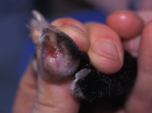
Digital metastasis of a primary lung tumour (Photo Arnaud Muller)
11.2 Feline pemphigus foliaceus
In feline pemphigus foliaceus, a creamy material covers the claw fold, together with inflammation. This paronychia with creamy to caseous exudate affecting several claws is specific to cats. Sampling is simple: the creamy, caseous material is scraped from around the claws, and cytology reveals acantholytic cells accompanied by non-degenerate neutrophils. Paronychia has been reported as a more frequent sign in cats than in dogs.
11.3 An onychopathy of unknown aetiology
Finally, Dr Zanna mentions a recent article on a cat in which the authors had great difficulty classifying what they observed. Histologically, they found acantholytic cells suggestive of pemphigus foliaceus; clinically, the picture was different, without the creamy, caseous material of the paronychia described above, and the lesions were more reminiscent of symmetrical lupoid onychitis. They finally settled on a diagnosis of generalised pustular onychopathy of unknown aetiology. The case is a reminder that the classification of some onychopathies remains open.
12 Decision summary by number of claws affected
In dogs, when a single claw is affected, the reasoning is simple: traumatic injury or tumour. When one or two claws are affected, onychomycosis is one of the best differential diagnoses. When many claws are affected in an otherwise healthy individual, symmetrical lupoid onychitis is one of the most important diagnoses.
When many claws are affected together with other signs, cutaneous or non-cutaneous, the dominant sign points the way. If paronychia dominates, think of bacterial paronychia, Malassezia overgrowth, and skin conditions that first affect the folded parts of the digits and can secondarily reach the claw fold, such as demodicosis. If onychogryphosis dominates, the two best differential diagnoses are leishmaniosis and ischaemic dermatopathy, bearing in mind that leishmaniosis may be the underlying disorder that induces ischaemic dermatopathy. If onychomadesis dominates, rare diseases must be considered: subepidermal blistering diseases, genodermatoses, or pemphigus in cats.
In cats, two entities are worth knowing for what they imply: lung-digit metastatic syndrome, in which the claw is sometimes the first sign, and pemphigus foliaceus, whose creamy paronychia is specific to the species.
Conclusion
The claws of dogs and cats provide a wealth of information. Claw disorders remain poorly documented and are usually treated as a side issue. Yet in many cases the claw is the indicator of a much wider dermatological or systemic disease, and recognising it early could improve clinical outcomes.
In practice, the claw is approached like any other dermatological condition, without fixating on it or ignoring the rest of the animal. Next, the number of claws affected is the first guide, one or two claws pointing to a local cause and several claws to a systemic one. Finally, speaking of symmetrical lupoid onychitis rather than onychodystrophy is a reminder that inflammation of the claw bed precedes the dystrophy it produces.
Zanna G. Claw diseases: what you need to know. Istituto Veterinario di Novara, Italy. ECVD-ESVD Meeting Bilbao 2025
