Alopecia areata (Pélade) is a rare autoimmune disease in domestic carnivores, characterized by non-scarring hair loss. This condition, well-documented in human medicine, remains insufficiently studied in veterinary dermatology, thus limiting knowledge relating to therapeutic protocols and prognosis in canine patients.
September 2025
The available clinical data concerning this pathology remain fragmented, making it necessary to deepen the understanding of its clinical manifestations, histopathological characteristics, and therapeutic management modalities.
Objectives and methodology of the study
This retrospective investigation revolves around the analysis of fourteen cases of canine alopecia areata, with the aim of characterizing clinical manifestations, histopathological features and evaluating the efficacy of various therapeutic approaches. Strictly defined inclusion criteria comprised the presence of leukotrichia and non-inflammatory alopecia, the absence of immunosuppressive treatment at the time of biopsy, histopathological confirmation of bulbite, and the availability of histological slides for review.
Case collection was carried out through archives of a private veterinary pathology laboratory and solicitations from specialized veterinary dermatology mailing lists. The analysis of medical records allowed documentation of the age of onset, duration of evolution before diagnosis, initial and progressive clinical characteristics, previous diagnostic explorations, concomitant pathologies, and instituted therapeutic modalities.
Epidemiological characteristics and clinical manifestations
Case details and demographic data
The analysis reveals a predominance of Labrador Retrievers and their crossbreeds, representing four of the fourteen cases studied. Purebreds account for most of the remaining cases, with a diversity including Basset Hounds, Golden Retrievers, Great Pyrenees, Boxers, German Shepherds, and French Bulldogs. The median age of onset for lesions is five years, ranging from six months to twelve years. The male-to-female ratio of 1.3 contrasts with human observations showing a female predominance.
Initial clinical presentation
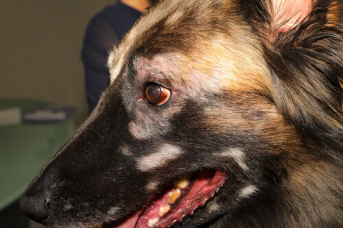
Initial clinical manifestations are primarily characterized by non-inflammatory alopecia, devoid of erythema, crusts, or excoriations in twelve patients. Pinnal and periocular leukotrichia constitute the other observed presentation modes. Anatomical distribution significantly favours the head region, affected in ten of the thirteen documented dogs, followed by the distal extremities and trunk. On the face, periocular and pinnal locations predominate, while post-auricular, pre-auricular, muzzle, chin, and dorsal skull areas can also be affected.
Alopecia areata in a German Shepherd
Concomitant pathologies and comorbidities
The prevalence of concomitant pruritus reaches seventy-nine percent of cases, with five dogs previously diagnosed with atopic dermatitis. This association suggests shared pathophysiological mechanisms or common predisposing factors. Two patients had histories of recurrent external otitis, while two others were under ophthalmological follow-up for uveitis with secondary glaucoma and corneal opacities with conjunctivitis.
Diagnostic explorations and therapeutic management
Diagnostic approach
Preliminary investigations before histopathological diagnosis included haematobiochemical tests for four dogs, revealing parameters within physiological limits. Thyroid exploration, performed in seven patients, did not reveal any abnormalities, with three dogs only benefiting from total thyroxine dosage. Parasitological examinations through skin scrapings and dermatophyte cultures were respectively negative in seven and three patients.
Therapeutic approaches and treatment modalities
Clinical evolution and prognosis
Clinical follow-up, available for twelve dogs over a period ranging from two months to four years, reveals a tendency to relapse upon reduction or discontinuation of immunosuppressive treatments. Five patients experienced recurrence of characteristic lesions during therapeutic withdrawal attempts. Spontaneous remission, observed in only two dogs, is less frequent than previously reported, suggesting a more reserved prognosis than initially described in the veterinary literature.
Distinctive histopathological characteristics
Quantitative analysis of hair follicles
Histopathological examination of forty-two skin samples reveals involvement of seventy-one percent of anagen hair bulbs. The severity of peribulbar cellular infiltration, graded according to the diameter of the inflammatory area, is distributed from minimal to moderate inflammation for twenty-eight percent of the affected bulbs, moderate for fifty-six percent, and severe for sixteen percent.
Composition of the inflammatory infiltrate
The peribulbar infiltrate presents a complex cellular composition dominated by lymphocytes, present in all patients. Plasma cells accompany this infiltration in thirteen dogs, while eosinophils, macrophages, and neutrophils complete the inflammatory picture respectively in seven, six, and six patients. The extension of inflammation to the follicular isthmus level, observed in fifty percent of cases, requires a differential diagnosis with pseudopelade.
Follicular architectural modifications
Follicular dysplasia, characterized by a dystrophic and deformed appearance of the lower portion of the hair follicle, affects ninety-three percent of patients, contrasting with the twenty-four percent reported in previous studies. This discrepancy could result from differences in biopsy techniques, case severity, or applied diagnostic criteria.
Follicular keratosis represents a universal histopathological characteristic in this series, observed in all patients with adequately represented sebaceous glands. This finding suggests that follicular keratosis could constitute a new diagnostic criterion for canine alopecia areata, akin to observations made in human medicine.
Other tissue modifications
Peribulbar pigmentary incontinence, characterized by the presence of large melanin granules in macrophage cytoplasm, affects seventy-nine percent of cases. This histological feature distinctly differs from the pigmentary incontinence observed in the uveodermatologic syndrome, which presents a fine granular distribution of melanin.
Perifollicular fibrosis centred on anagen bulbs and mucinosis accompany nine and five cases respectively. The peri-vascular to interstitial dermatitis, of variable intensity ranging from traces to moderate, completes the histopathological picture in eight patients.
Therapeutic implications and future perspectives
Efficacy of Janus kinase inhibitors
The demonstrated efficacy of oclacitinib in this series aligns with the therapeutic advancements observed in human medicine with JAK inhibitors. Although the precise pathophysiological mechanisms of canine alopecia areata remain incompletely elucidated, the efficacy of oclacitinib suggests involvement of JAK-dependent signalling pathways, notably those involving gamma interferon, interleukin-2, and interleukin-15.
Long-term management and prevention of relapses
The marked tendency for relapses during the reduction of immunosuppressant treatments underscores the chronic nature of this condition. This characteristic necessitates careful consideration of maintenance therapy strategies and the search for a balance between clinical efficacy and potential adverse effects. Despite its demonstrated efficacy, cyclosporine may cause side effects limiting its prolonged use, such as gingival hyperplasia observed in one patient from this series.
Complementary diagnostic tools
Trichoscopy, widely used in human medicine for the diagnosis and follow-up of alopecia areata, remains underutilized in veterinary practice. This technique could prove particularly useful for identifying characteristic signs such as obstructed follicles and for monitoring therapeutic response.
Study limitations and research perspectives
The limited size of the sample, inherent to the rarity of this condition, constitutes a major constraint for extrapolating the results. The retrospective nature of the study and the variability in follow-up methods between patients also limit the scope of the conclusions. Controlled prospective investigations are necessary to refine optimal therapeutic protocols and determine prognostic factors.
The absence of investigations into the pathophysiology of canine alopecia areata contrasts with the considerable advances made in human medicine. Transcriptomic and immunohistochemical studies could elucidate the underlying mechanisms and identify new therapeutic targets.
This study significantly enriches the knowledge on canine alopecia areata by documenting fourteen new cases and identifying follicular keratosis as a consistent histopathological feature. The results suggest a more reserved prognosis than previously reported, with a marked tendency for relapses requiring prolonged maintenance treatments. The demonstrated efficacy of oclacitinib opens promising therapeutic perspectives, although further studies are needed to optimise its use in this indication.
Mathai M, Banovic F, Thompson L, Trainor K. Canine alopecia areata: a retrospective study of clinical, histopathological features and treatments in 14 dogs. Vet Dermatol. 2025;0:1-10.
