Dermatozoonoses constitute a diverse group of skin conditions transmissible between vertebrate animals and humans. These pathologies, although relatively rare compared to all known zoonoses, represent a significant health challenge, particularly in urban contexts where proximity to pets is intensifying.
The veterinary practitioner, at the interface of animal and human health, plays a crucial role in the detection, treatment, and prevention of these conditions.
The close relationship between humans and animals, while offering undeniable psychological and social benefits, also exposes pet owners and professionals in contact with animals to various transmissible pathogens. These dermatozoonoses, of mycotic, parasitic, bacterial, or viral origin, require particular vigilance and a collaborative approach between medical doctors and veterinarians for optimal management of patients, whether two-legged or four-legged.
Definition and Classification of Dermatozoonoses
Dermatozoonoses represent a specific subset of the vast field of zoonoses. They are defined as diseases or infections naturally transmissible from vertebrate animals to humans, and vice versa, clinically manifesting in humans as skin lesions, with the exception of generalized allergic reactions. This term derives from the Greek roots “zoo” (animal) and “nosos” (disease), conceptualized by Virchow in the 19th century, and later clarified by the World Health Organization in 1959.
In urban environments, the main vertebrates responsible for these conditions are pets – domestic carnivores like dogs and cats, but also rodents and lagomorphs increasingly present in our homes. Exposed populations include not only pet owners, but also various at-risk professions: veterinarians and their staff, breeders, groomers, and other professionals in regular contact with these animals.
Classification by Transmission Modalities
The World Health Organization proposes a classification of zoonoses into four distinct categories, based on their transmission modalities:
- Orthozoonosis or Direct Zoonosis: The causal agent requires only one vertebrate species for its maintenance, although it can affect several species. This species allows transmission to humans. The majority of classic infectious zoonoses like rabies, anthrax, or brucellosis belong to this category.
- Cyclozoonosis: In this case, the biological cycle involves several vertebrate species, but only one of them is responsible for human contamination. Echinococcosis perfectly illustrates this process, with its cycle involving dogs and herbivores, where the dog acts as a contaminator of humans.
- Metazoonosis: This category requires passage through an invertebrate, usually an arthropod, which allows transmission to humans. Arboviruses like yellow fever (maintained in monkeys and marsupials then transmitted to humans by a mosquito), rickettsioses, and leishmaniasis fall into this classification.
- Saprozoonosis: These conditions require the passage of the causal agent into the external environment. Fascioliasis illustrates this mechanism, with the maturation of cercariae responsible for furcocercarial dermatitis.
This systematic classification allows for a better understanding of epidemiological cycles and, consequently, promotes the implementation of prevention strategies adapted to each type of transmission.
Mycotic Dermatozoonoses
Fungal infections transmissible between animals and humans constitute an important chapter of dermatozoonoses. These conditions, mainly represented by dermatophytosis and sporotrichosis, deserve particular attention due to their prevalence and potential impact on human health.
Dermatophytosis
Dermatophytosis, commonly known as “ringworm,” represents superficial infectious mycoses caused by epidermotropic, keratinophilic, and keratinolytic fungi – dermatophytes. These fungi have the particularity of feeding on keratin, a protein constituting superficial structures like hair, fur, nails, and the stratum corneum of the epidermis.
Zoophilic dermatophytes, of animal origin, constitute a major source of dermatozoonoses. Three main species are to be considered:
- Microsporum canis: The main reservoir is the cat (more rarely the dog, especially the Yorkshire Terrier breed, and other small mammals). This species is responsible for about 95% of feline dermatophytosis and 65% of canine cases.
- Trichophyton mentagrophytes: Its reservoir consists of rodents, lagomorphs, and equids. The percentage of asymptomatic carriers varies considerably depending on the species: approximately 15% in guinea pigs, 10 to 40% in rabbits, and nearly 50% in mice and rats.
- Trichophyton verrucosum: Primarily found in ruminants.
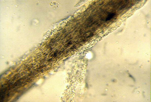
Ringworm hair
Most dermatophytes show a close adaptation to a target species, in which they generally cause few clinical signs. In cats, for example, the incidence of asymptomatic M. canis carriers is around 10%, but can reach much higher percentages in stray cats or certain breeds like Persians. Alarmingly, about 50% of people in contact with an infected cat, whether an asymptomatic carrier or clinically ill, develop skin lesions.
Clinical Presentation in Animals
In cats, classic M. canis lesions are characterized by single or multicentric, erythematous, non-pruritic, scaly, centrifugally spreading hair loss. Preferred locations are the face, ears, and paw extremities. A late diagnosis may reveal loco-regional or generalized alopecia, sometimes accompanied by pruritic inflammation. In some cases, only a seborrheic keratitis with scaling along the back is observed. Onychomycosis due to M. canis is rare, while mycetomas occur exclusively in immunocompromised individuals and in the Persian breed.
In dogs, in addition to M. canis, three other species are found: T. mentagrophytes, M. persicolor (both zoophilic agents), and M. gypseum (geophilic agent). Certain breed predispositions exist: Yorkshire Terriers seem particularly vulnerable to generalized ringworm by M. canis, while terriers (Fox Terrier and other hunting dogs) show increased susceptibility to facial dermatophytosis caused by T. mentagrophytes and M. persicolor.
The majority of affected dogs present classic lesions: unique or multicentric, rounded, slightly scaly hair loss, centrifugally developing, generally not very itchy. Preferred locations are the face and the distal extremities of the limbs. Kerions (inflammatory nodular suppurative lesions) are regularly observed, particularly with T. mentagrophytes.
In rodents and lagomorphs, increasingly popular as pets, the almost exclusive agent is T. mentagrophytes, more rarely M. canis. Clinically, dermatophytosis manifests as centrifugal hair loss, often pruritic and very inflammatory.
Diagnosis of Dermatophytosis
Diagnosis relies on a multi-faceted approach:
- Wood’s lamp examination: A typical green fluorescence of contaminated hairs is observable in 50% of M. canis cases. This technique nonetheless requires some experience to distinguish specific fluorescence from artifactual fluorescence.
- Direct microscopic examination of hairs and scales: It can reveal an alteration of the hair shaft, sheathed by spore sleeves (endoectothrix type hair invasion in the case of M. canis).
- Mycological culture: It allows for definitive identification of the responsible dermatophyte. In asymptomatic carriers, the sample can be taken by brushing the entire body surface with a sterile toothbrush or a sterile carpet square, then cultured on Sabouraud agar.
Treatment and Prevention
Treatment is imperative, even if spontaneous healing is possible (although it may take several months, or even years). Therapeutic efficacy depends on taking into account two essential factors:
- The high contagiousness of dermatophytosis
- The prolonged resistance of spores in the environment (several years)
The therapeutic approach combines:
- Topical treatment: clipping (discussed on a case-by-case basis), shampoos and baths based on enilconazole or miconazole combined with chlorhexidine
- Systemic treatment: griseofulvin, ketoconazole, itraconazole
- Environmental treatment: enilconazole or bleach
Treatment should be continued until mycological cure confirmed by negative cultures, with two negative controls spaced one month apart. In breeding facilities, the major problem remains permanent recontamination due to the significant spore load in the environment, making eradication practically impossible despite sanitary fallow periods. Ideally, in uninfected breeding facilities, any new animal should be quarantined until a negative mycological culture is obtained.
Impact on humans
In humans, dermatophytosis of animal origin is characterized by remarkable clinical polymorphism:
- Erythematous-squamous lesions of the scalp (mainly due to M. canis)
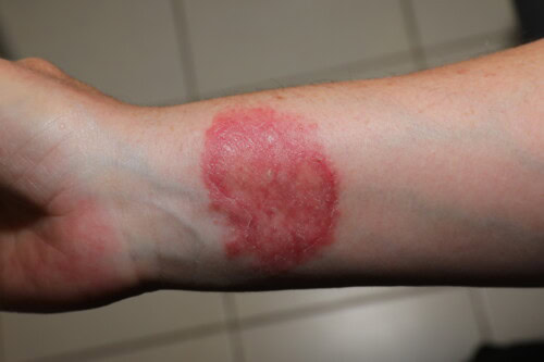
- Circular erythematous-squamous lesions of glabrous areas, often polycyclic with a vesiculosquamous border (M. canis, T. mentagrophytes)
- Crusted and painful lesions with severe inflammatory reaction (kerion) of the scalp, neck or beard (sycosis), mainly caused by T. mentagrophytes, more rarely M. canis
Human contamination during ringworm
It is important to note that ringworm is listed in table no. 46 of occupational diseases (cutaneous mycoses). According to the Labor Code and European directives, employer obligations include the prevention of occupational risks, specific regulatory measures, hygiene and safety measures, and medical monitoring.
Sporotrichosis
Sporotrichosis represents a deep mycosis with high zoonotic potential, caused by a dimorphic fungus, Sporothrix schenckii. This pathogen has a remarkable characteristic: it exists in filamentous form in the surrounding environment (decomposing plants, humus, soil) and transforms into yeast after penetrating host tissues through a skin breach.
Modes of contamination
While punctures and wounds from contaminated plant elements constitute the classic mode of contamination, the infected cat represents a particularly worrying source of contamination. Indeed, pet owners, veterinarians, and their staff are exposed to a significant risk through contact between their pre-existing skin lesions and exudates during manipulation of the sick animal.
A distinctive feature of this infection is the difference in fungal load depending on the host species: in dogs or humans, the fungal agent is present in limited quantities in lesions, whereas in cats, fungal elements reach extremely high concentrations. Transmission to humans mainly occurs through skin trauma: bite or scratch from a contaminated cat.
It should be noted that canine sporotrichosis, rarer than its feline counterpart, has not yet been associated with proven cases of human contamination.
Clinical presentation in animals
In dogs and cats, three distinct clinical forms have been described, with a variable incubation period from one week to two months:
- Cutaneous-lymphatic form (80% of cases): It is characterized at the inoculation site by the progressive development of a single nodule, initially asymptomatic then ulcerated, located on the face or a paw extremity. Other ulcerated and fistulated nodules may appear along the lymphatic vessels.
- Strictly cutaneous form (less frequent): Preferred locations are the paw extremities, which present nodular lesions or areas of hair loss with raised borders, ulcerated and crusted.
- Generalized form: Observed mainly in immunocompromised cats, it results from the hematogenous dissemination of infective spores.
Diagnosis and Treatment
Diagnosis relies on cytological examination of skin smears and biopsies, which reveal a remarkable abundance of fungal elements in cats. Mycological culture confirms the diagnosis.
Treatment is based on the administration of systemic antifungals, with a preference for itraconazole and fluconazole. In systemic forms, amphotericin B yields excellent results. Treatment should be continued for at least one month after apparent clinical resolution. In cats, in addition to systemic fungicide treatment, strict hygienic measures are essential:
- Careful cleaning of hands and arms with antifungal substances (chlorhexidine, povidone-iodine)
- Mandatory wearing of protective gloves
- Clear information for owners regarding the major zoonotic risk
Due to the high risk of transmission and the prolonged duration of treatment, euthanasia may be legitimately considered for carrier cats.
Manifestations in humans
In humans, incubation varies from three weeks to three months. The cutaneous-lymphatic form, the most frequent, manifests at the inoculation site as a progressive nodule, initially asymptomatic then ulcerated, typically located on the back of the hand, a finger, a foot, or the face.
Parasitic Dermatozoonoses
Cutaneous conditions of parasitic origin transmissible between animals and humans constitute a heterogeneous but important group of dermatozoonoses. These parasitoses, involving various mites, insects, or helminths, present varied clinical pictures in both animals and humans.
Mange
Sarcoptic Mange
Sarcoptic mange represents a highly contagious acariasis due to the proliferation in the epidermis of mites from the Sarcoptidae family, Sarcoptes scabiei var. canis. Its importance in veterinary and human dermatology stems from its increasing frequency, clinical severity, and proven zoonotic potential.
The species primarily affected by Sarcoptes scabiei var. canis are dogs, foxes, and ferrets, but also occasionally cats, humans, and horses. The evolutionary cycle is characterized by its rapidity (10 to 13 days) under favorable environmental conditions. Females, extremely prolific, lay 2 to 3 eggs daily (about 50 eggs per female) for 2 to 4 weeks. After fertilization, they dig epidermal burrows (unlike the tunnels observed with human sarcoptes) to deposit their eggs.
These eggs hatch in 2-3 days, releasing hexapod larvae that develop into nymphs and then adults. Sarcoptes feed on epidermal tissues (histophagous). Their survival in the external environment is limited to about 10 days, requiring specific conditions (15-25°C, relative humidity between 25 and 85%).
Sarcoptic mange remains underdiagnosed in dogs. It primarily affects young dogs under one year of age, especially in group settings (kennels, shelters), but can also affect adult or older dogs weakened by an intercurrent illness.
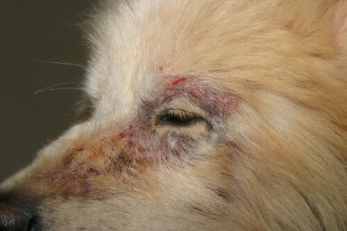
Sarcoptic mange with facial involvement
The pathogenic power of Sarcoptes scabiei var. canis is exerted through different mechanisms:
- Mechanical and chemical actions (inoculation of vasodilatory and anticoagulant proteins)
- Antigenic action (excrement, molting products, saliva)
- Induction of hypersensitivity phenomena of types I, IV and III
Type III hypersensitivity can cause immune complex deposits in various organs, particularly the kidneys, causing glomerulonephritis. This systemic dimension justifies considering canine sarcoptic mange as a general disease and not only dermatological.
Clinically, after a variable incubation (around 3 weeks post-contact), the classic picture combines:
- Intense pruritus with positive otic-pedal reflex
- Erythematous and papular lesions (“mange pimples”)
- Alopecia in patches
- Crust formation
The lesion distribution is characteristic at the beginning of the evolution, primarily affecting the face (free edges of the ear flaps), limbs (elbows), and sternum. Systemic manifestations can occur during old infestations or in older dogs: hyperthermia, anorexia, weight loss, polyuria-polydipsia secondary to immunological glomerulonephritis.
Atypical forms are increasingly reported:
- Frustrating localized forms, not very itchy and not very contagious
- “Norwegian mange” characterized by thick compact scales, moderate pruritus, and the massive presence of sarcoptes at different evolutionary stages in skin scrapings (typically in immunocompromised animals)
The definitive diagnosis is sometimes difficult. Microscopic examination of deep skin scrapings, carried out in predilection areas, reveals parasites or their traces (sarcoptes, eggs, droppings) in only about 50% of cases. Serological diagnosis is an alternative, with a sensitivity and specificity of around 80-90%. Faced with a strong clinical suspicion without parasitological confirmation, a trial treatment is recommended according to the principle “if you suspect it, treat it”.
Treatment involves two approaches:
- Topical acaricides (diluted amitraz)
- Systemic acaricides as spot-on (selamectin or moxidectin)
The zoonotic dimension is significant, with human contaminations observed in 25 to 50% of canine sarcoptic mange cases. Incubation in humans is 8 to 15 days, resulting in prurigo of the trunk, arms, and legs. Characteristically, no scabies burrow is observed, unlike human scabies.
This is explained by the fact that Sarcoptes scabiei var. canis cannot survive more than 15 to 20 days in humans, due to strict host specificity. The parasite is unable to reproduce in human skin (absence of ovigerous females in the epidermis, no burrows) and remains confined to the surface without burrowing or antigenic action.
Therefore, appropriate treatment of the mangy animal is usually sufficient to resolve symptoms in humans – sarcoptic mange being considered a “hemizoonosis” where the parasite dies quickly in human skin without reproducing. Persistent human symptoms should raise suspicion of a persistent source of contamination: untreated or poorly treated animal, asymptomatic congener not identified, or parasite survival in the environment.
Notoedric Mange
Notoedric mange is a contagious acariasis primarily affecting cats, rats, and hamsters. It is caused by the multiplication on the surface and in the epidermis of psoroptic mites from the Sarcoptidae family: Notoedres cati in cats and Notoedres muris in rodents.
Relatively rare in cats in mainland France, this condition is more common in overseas departments and territories (Reunion Island, Antilles), as well as in Italy, Slovenia, and Spain. In hamsters and rats, it is one of the most common pruritic dermatoses.
The biological cycle of notoedres is comparable to that of sarcoptes, with marked contagiousness, particularly by direct contact, potentially affecting cats, dogs, and humans. This infestation can occur enzootically or epizootically with significant morbidity. Susceptibility factors include young age (kittens), immunosuppression (FeLV or FIV positive cats), and, in rodents, pup or pregnant female status.
Clinically, in cats, lesions generally begin on the face (nose bridge, lips, eyelids, ear flaps) before generalizing to the limbs and perianal and abdominal regions. They manifest as diffuse erythematous and scaly hair loss, rapidly progressing to crust formations. Pruritus is usually intense.
In hamsters, cutaneous manifestations include crusted lesions localized preferentially to the muzzle, ear flaps, and limb extremities, with frequent genital involvement. Pruritus varies from moderate to intense.
In rats, the lesion distribution is more restricted, limited to the free edge of the ear flaps and the muzzle, in the form of pseudo-tumoral warts. Papulo-crusted lesions are generally observed on the tail, but generalization is unusual. The intensity of pruritus varies from moderate to severe.
Human contaminations, whose precise incidence is difficult to evaluate, produce skin signs similar to those of canine sarcoptic mange: prurigo of the trunk, arms, and legs. As with Sarcoptes scabiei var. canis, notoedres cannot reproduce in human skin (absence of epidermal ovigerous females and burrows) and remain confined to the surface, without burrowing action or significant antigenic response. The resolution of mange in the animal therefore generally leads to healing in humans, this notoedric mange also being considered a hemizoonosis.
Diagnosis, relatively simple, relies on the identification of notoedres at various evolutionary stages (adults, nymphs, larvae, eggs) and their droppings during skin scrapings.
Treatment involves systemic acaricides (avermectins and milbemycins), with the need to treat all animals in the group, whether or not they show clinical signs.
Trixacarus Mange
Mange due to Trixacarus caviae is a contagious acariasis specific to guinea pigs (and occasionally mice), caused by a psoroptic mite of the Sarcoptidae family. The triggering factors remain poorly understood, with often subtle contamination conditions and a variable incubation period.
Reconstructing the history of infestation is often complex, as contamination frequently precedes the acquisition of animals. These guinea pigs likely harbor a small number of parasites without clinical manifestation, until environmental changes (food, habitat, overcrowding) or an alteration of their health status cause parasitic multiplication beyond the pathogenic threshold.
Notably, the presence of severely affected animals does not systematically lead to the contamination of congeners in contact, suggesting an individual component in susceptibility to infestation.
The clinical picture associates constant, early, and often intense pruritus with rapidly generalized lesions: erythema, papules, scales, progressing towards extensive crust formations. The distribution can be loco-regional or generalized. The general condition can deteriorate during chronic evolution, with apathy, anorexia, weight loss, and sometimes a fatal outcome.
Human contaminations, regularly reported, result from frequent and prolonged contact with the sick animal. Infestation episodes have notably been described in school communities, involving kindergarten children in contact with a severely affected guinea pig.
In humans, this mange manifests as a pruritic papular dermatosis (prurigo type) mainly affecting the arms, neck, and legs. As with other animal mangy, appropriate treatment of the animal is usually sufficient to resolve human symptoms, this trixacarus mange also being a hemizoonosis where the parasite cannot reproduce in human skin.
The therapeutic protocol relies on the use of topical or systemic acaricides (avermectins or milbemycins), with mandatory treatment of all congeners. Cleaning and acaricide treatment of the environment complete the management.
Cheyletiellosis
Cheyletiellosis represents a set of parasitic dermatoses caused by mites of the genus Cheyletiella, belonging to the Cheyletidae family. Three main species are identified, each with a host preference: Cheyletiella yasguri (dog), Cheyletiella blakei (cat), and Cheyletiella parasitivorax (rabbit).
These mites have particular biological characteristics: adults lay eggs at the base of the hairs and feed on skin debris and tissue fluids. Unlike other surface ectoparasites, cheyletiella can burrow into epidermal debris, or even into the stratum corneum, forming pockets. Their mobility on the skin is remarkable.
They are obligate parasites completing their entire life cycle on their host, with a development duration of approximately 35 days. Transmission primarily occurs through direct contact with clinically affected or asymptomatic carrier animals, but also indirectly via the environment, where females can survive up to 10 days. Interspecific contaminations are possible, and survival could be prolonged under favorable environmental conditions (relatively low temperature, high humidity, moderate light). The parasites can contaminate animal bedding, wall and floorboard crevices, sometimes even in the absence of animals.
Cheyletiellosis primarily affects young animals (puppies and kittens from kennels or catteries), but also adult dogs (often asymptomatic carriers) and adult cats. Certain breed predispositions have been observed: dwarf canine breeds (Yorkshire Terrier, Bichon, Poodle) and, in cats, the Persian breed.
The clinical presentation varies depending on the species and age:
- In puppies: intense pruritus with positive otic-pedal reflex and pronounced scaling affecting the head, back, and loins
- In kittens: discrete signs limited to pityriasis-like dorsal-lumbar scaling
- In adult cats: more inflammatory skin lesions with pruritic papulo-crusted dermatitis
- In rabbits: often asymptomatic infestation or pruritic and scaly dermatosis mainly affecting the trunk
Human contamination is frequent (>50% of cases), generally occurring from C. blakei and C. yasguri. This often underestimated transmission mainly results from direct contact with the parasitized animal (clinically affected or asymptomatic carrier), but can also occur indirectly.
In humans, clinical manifestations appear rapidly, from the second day after contact, in the form of very itchy papules localized on the forearms, elbow creases, arms, chest, abdomen, and thighs.
Diagnosis, generally straightforward, relies on the identification of cheyletielles by skin scrapings, brushing, or “scotch test” and microscopic observation of scales and debris. Visualization of parasites is usually easy in dogs (adults, nymphs, eggs) but more difficult in cats, where adults are rarely identified and only eggs at the base of the hairs can be observed. In rabbits, parasitic forms are easily detectable.
Specific treatment combines:
- Topical or systemic acaricides (avermectins or milbemycins)
- Prolonged treatment (minimum eight weeks) due to the resistance of eggs to acaricides
- Mandatory treatment of all animals in contact
- Environmental treatment
Isolation and appropriate treatment of animals generally lead to spontaneous resolution of lesions in humans, as cheyletielles cannot reproduce in human skin.
Pulicosis
Flea infestations (pulicosis) are among the most common ectoparasitoses in dogs and cats. The predominant species is Ctenocephalides felis felis – “the cat flea” – more rarely Ctenocephalides canis. Although pulicosis is not strictly considered a zoonosis in the proper sense, their frequency and harmful consequences in humans justify their inclusion in this analysis of dermatozoonoses.
Fleas are wingless (aphaniptères), laterally flattened insects. Adults, cosmopolitan and sedentary parasites, spend most of their time on the host animal. Their biological cycle has several specificities: females begin laying eggs on the host within 24 to 48 hours after their first blood meal. These eggs, with a smooth surface, fall into the environment where development occurs (eggs, three larval stages, nymphal stage).
The complete cycle takes approximately three weeks under optimal conditions, but can be slowed down under unfavorable environmental conditions. A crucial element of epidemiology is the ability of pre-emerged adults (still in nymphal cocoons) to persist for several months in the environment, constituting a considerable parasitic reservoir. These adults emerge under the influence of specific stimuli (vibrations, light, chemical signals) and form what is commonly known as “floor fleas.”
Flea infestation is generally inapparent in healthy animals. Healthy dogs experience moderate parasitic pressure, and the flea population is naturally limited by self-grooming behaviors (mouthing, licking) and scratching movements that expel parasites from the coat. This particularity explains the occasional difficulty in detecting parasites on infested animals.
The pathological consequences of pulicosis have two dimensions:
- Mechanical skin irritation due to repeated bites and parasite movements
- The development, in certain sensitized subjects, of hypersensitivity dermatitis to flea bites (FAD), which is the most common allergic dermatosis in both cats and dogs
In dogs, FAD manifests as dorsal-lumbar alopecia combining erythema, papules, and crusts, with generally intense pruritus. In the absence of adequate treatment, secondary infectious complications are frequent. In cats, the picture is characterized by pruritic papulo-crusted dermatitis, eosinophilic plaques, and/or dorsal-lumbar alopecia.
Transmission to humans primarily occurs during massive infestations, when overcrowding forces fleas to change hosts. It should be noted that direct transmission of adult fleas between animals or to humans is relatively limited (10 to 15% on average). Human contamination mainly comes from freshly emerged young adult fleas from cocoons, which actively seek an available host, whatever it may be.
In humans, flea allergy dermatitis preferentially affects the limbs (ankles, wrists) and, in children, can extend to the trunk. The intensely pruritic lesions appear as urticarial papules, wheals, or transient vesicles. Impetigo secondary to scratching and bacterial superinfections is sometimes observed.
The diagnosis of pulicosis relies on the direct identification of adult fleas or their droppings, either with the naked eye or by brushing with a specific flea comb. For FAD, diagnosis is based on suggestive anamnestic elements (seasonal dermatosis, presence of several dogs/cats in the environment) and compatible clinical lesions (pruritic dorsal-lumbar dermatosis). Paradoxically, in animals with FAD, visible flea infestation is often minimal.
In dogs, intradermal tests using total flea extracts can confirm FAD by positive immediate reactions at 20 minutes and/or delayed reactions at 48 hours, although their absence does not exclude the diagnosis. These tests are considered unreliable in cats.
Treatment of pulicosis and FAD should never be trivialized and requires a comprehensive approach:
- Elimination of fleas on the affected animal
- Mandatory treatment of congener animals
- Environmental sanitation
This integrated strategy often requires close collaboration with the owner to establish an effective control program. Flea control on the animal relies on the use of residual adulticides, while the management of non-parasitic stages in the environment involves insect growth inhibitors and environmental adulticides. Complementary mechanical measures (methodical cleaning, elimination of ecological niches) are also essential.
Helminthic Dermatozoonoses
Cutaneous Larva Migrans
The phenomenon of cutaneous larva migrans (CLM) is observed mainly in mainland France in people returning from tropical regions, where stray dogs contaminate beaches with their excrement. These dogs, usually unmedicalized and dewormed, are frequently infested with hookworms (Ancylostoma spp. and Uncinaria spp.), digestive nematodes responsible for hemorrhagic gastroenteritis in them. However, the recent description of an enzootic larva migrans in Brittany calls for increased vigilance on mainland territory.
The cutaneous manifestations are linked to the transcutaneous penetration of infective L3 larvae. Clinically, they result in papules, often crusted and pruritic, sometimes pustular, preferentially located on thin-skinned areas (abdomen) and limbs (interdigital spaces and palmar surfaces). These signs, relatively non-specific, require a precise anamnesis suggesting potential contact with contaminated environments (dog living in a kennel, rural area, hunting dog).
Beyond cutaneous manifestations, infestation can cause respiratory signs related to larval migration (bronchopneumonia), often underdiagnosed. Digestive disorders (hemorrhagic enteritis) and general signs (weight loss, anemia) are regularly observed during chronic infestations.
Sources of contamination include carrier dogs (and cats) as well as moist soils contaminated by infective L3 larvae. Poorly maintained kennels with dirt floors constitute an ideal environment for larval development, explaining the particular vulnerability of hunting dogs. Also noteworthy is the possibility of indirect infestation by dogs ingesting small rodents that have themselves ingested L3 larvae.
Diagnosis of transcutaneous migrations is often difficult. Direct detection of L3 larvae by skin scrapings is rarely conclusive. Histopathological examination of skin biopsies may suggest these migrations (eosinophilic infiltrate and occasionally presence of larvae). Coproscopic examination generally allows easy identification of eggs, often numerous.
Prevention and control involve a multifaceted approach:
- Destruction of contaminated environments
- Regular and reasoned deworming, particularly of pregnant bitches (larvicidal anthelmintics)
- Reconstruction of infested dirt kennels
- Daily collection of excrement
- Intensive weekly cleaning with cresol
In humans, the larva penetrating the skin produces a characteristic serpiginous eruption. However, this infestation constitutes a parasitic dead end, with lesions generally regressing spontaneously within a few weeks to a few months.
Furcocercarial Dermatosis or Swimmer’s Itch
Furcocercarial dermatosis, also known as swimmer’s itch, represents an increasingly common seasonal dermatozoonosis (June to September). This metazoonosis is caused by the epidermal penetration of larvae of a trematode, Trichobilharzia ocellata, a duck parasite, sometimes improperly called “duck flea”. Contamination occurs during freshwater swimming, particularly in lake areas (Lake Annecy, Lake Bourget, Lake Geneva, Swiss and Italian lakes).
The parasitic cycle involves the trematode T. ocellata, a digestive parasite of ducks, excreted in feces. These trematodes are then ingested by aquatic snails of the genus Lymnaea, notably Lymnaea stagnalis. Cercariae (larval forms) are subsequently released into the water where they can contaminate ducks (definitive hosts) as well as humans or dogs (accidental hosts) during swimming.
Although dogs can present lesions similar to those observed in humans, it is important to emphasize that in no case can humans become contaminated from an affected dog.
In humans, the clinical picture is characterized by an intensely pruritic, maculo-papular eruptive dermatitis, localized to exposed areas. The sudden appearance of papules generally occurs within 10 to 30 minutes following immersion in fresh water. The evolution is favorable, with spontaneous healing in 2 to 3 weeks, although symptomatic treatment (antihistamines or topical corticosteroids) is frequently necessary.
Sanitary prophylaxis relies on interrupting the parasitic life cycle, involving the elimination of snails and ducks from affected water bodies. As eliminating molluscs is particularly difficult, bathers are advised to avoid shallow water areas rich in aquatic vegetation, a preferred habitat for snails.
Leishmaniasis
Leishmaniasis is an infectious protozoonosis transmitted by inoculation, characterized by the intracellular multiplication of a flagellated protozoan, Leishmania infantum, in the cells of the mononuclear phagocyte system. Its transmission occurs through the bite of sandflies, with the dog as the main reservoir. In France, canine enzootic foci are concentrated mainly in the southeast, around the Mediterranean coast, from the Italian border to the Spanish border, and from sea level up to approximately 800 meters in altitude.
Although the parasite has also been isolated in foxes and cats, their epidemiological role seems marginal. Recent discoveries have also highlighted other modes of transmission: sharing of needles among heroin addicts, contamination by blood products, suggesting the occasional existence of an anthropozoonotic cycle.
In dogs, leishmaniasis presents as a general disease with remarkable clinical polymorphism. Epidemiological surveys conducted in the Alpes-Maritimes and Marseille region indicate that approximately one in two leishmaniotic dogs is an asymptomatic carrier. Although systemic in this species, the disease mainly manifests with skin lesions, never isolated but associated with various clinical signs:
- Polyadenomegaly (frequent)
- Splenomegaly (rarer)
- Alteration of general condition (asthenia, facial muscle atrophy)
- Various complications: bilateral uveitis, arthritis, glomerulonephritis (sometimes the sole clinical manifestation, with unfavorable prognosis)
Cutaneous lesions, typically chronic in evolution, exhibit great morphological diversity:
- Generalized exfoliative dermatosis affecting the head, ear flaps, and limbs
- Ulcerations of the paw extremities and pressure points
- Depigmentation of the nose (primary or secondary to ulcers)
- Thickening of the nose and/or paw pads
- Non-ulcerated nodules, single or multiple (particularly in certain breeds like the Boxer or Doberman)
- Generalized sterile pustular dermatitis
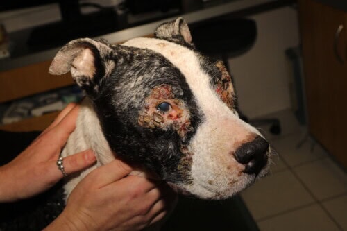
Canine leishmaniasis
In humans, leishmaniasis presents as a systemic disease primarily affecting children and immunocompromised adults. It can also manifest in a strictly cutaneous form, with lesions localized at the inoculation site, generally on exposed areas. These nodular, ulcerated, and crusted lesions are characteristically painless, of variable size, and chronic in evolution.
Diagnosis in veterinary medicine relies on various techniques:
- Direct parasite identification by cytology (adenogram, myelogram)
- Serology (ELISA, indirect immunofluorescence)
- Gene amplification (PCR)
In enzootic areas, systematic annual screening is recommended, ideally performed after the exposure season (November to January), given the variable but generally several-month incubation period.
The management of infected dogs raises many controversies. Indeed, even when treated, these animals remain carriers of the parasite. Human visceral leishmaniasis being potentially fatal in the absence of treatment, with an increasing incidence in enzootic areas, the elimination of the parasitic reservoir seems logical. However, this approach would penalize responsible owners of properly medicalized animals, while leaving an uncontrolled canine population. Paradoxically, systematic euthanasia of carriers, when applied, has not yielded the expected results and has even been accompanied by an increase in human cases.
The veterinarian plays a decisive role in informing owners about the risks and the need for rigorous monitoring, both clinical and biological.
Treatment of canine leishmaniasis is based on the combination of stibial derivatives and allopurinol, with close therapeutic monitoring. Despite owner awareness, a fraction of dogs inevitably escape control or receive intermittent self-medication, raising the serious problem of the potential emergence of resistant strains. Therefore, veterinarians should refrain from using certain highly effective molecules such as amphotericin B, which should be reserved exclusively for human medicine for this indication.
The development of a canine vaccine would represent the ideal solution for epidemiological control, but its development currently faces many obstacles.
Bacterial Dermatozoonoses
Benign Lymphoreticulosis of Inoculation or Cat Scratch Disease
Benign lymphoreticulosis of inoculation, more commonly known as cat scratch disease (CSD) due to its predominant mode of transmission, constitutes a subacute regional lymphadenopathy of bacterial origin in humans. The causative agent, Bartonella henselae (Bartonellaceae family), was only identified in 1992. Some cases could also be attributed to Bartonella clarridgeiae. The two known genotypes (I and II) of B. henselae are involved in this condition.
B. henselae is also associated, with B. quintana, with the etiology of bacillary angiomatosis and peliosis, vasculo-proliferative diseases observed mainly in HIV-infected patients.
The cat represents the main, if not the sole, reservoir of the bacterium. The dog’s role in carrying the infection seems very limited. Although rare, cases have been reported in the absence of direct exposure to an animal, suggesting other possible modes of transmission (flea or tick bites). Human contamination occurs in 70% of cases after a scratch and in 10% after a feline bite. Exceptionally, simple contact (cuddling, kissing) could allow contamination of a pre-existing skin or mucous wound, as illustrated by the oculo-glandular form sometimes observed in people who have probably rubbed their eye after stroking a cat.
Experimental infection in cats rapidly causes (less than one week) prolonged asymptomatic bacteremia, persisting for 2 to 3 months or more in some subjects (persistent recurrent bacteremia has been observed in one cat for 22 months). Some cats show remarkably high levels of bacteremia (greater than 10^6 CFU/ml of blood). Bacteremia is statistically more frequent in young cats (less than one year old). B. henselae and B. clarridgeiae can co-infect the same animal. Recently, two new species of Bartonella, B. koehlerae and B. weissii, have been isolated from cats in the United States, but their pathogenic role in CSD remains to be demonstrated.
Epidemiological studies indicate that a substantial proportion of tested cats are bacteremic, with a higher percentage among stray cats compared to domestic cats. Various surveys have revealed bacteremic cat rates ranging from 16.5% to 53% among stray feline populations, about one-third of which were infected with B. clarridgeiae.
The cat flea (Ctenocephalides felis felis) plays a predominant role in transmitting the infection within this species. B. henselae can also be isolated from fleas collected from bacteremic cats. The flea would eliminate the bacterium in its feces, thus contaminating the animal’s coat. Bacteria can multiply in the insect’s digestive tract and survive in its feces. The cat contaminates its claws during grooming, thus establishing the chain of transmission to humans. Stray cats, more frequently infected, represent a source of contamination for domestic cats.
CSD is a ubiquitous disease (estimated annual 22,000 human cases in the United States and 2,000 in the Netherlands in 2003) that can affect all ages, but primarily affects children and young adults. Half of the cases are reported in children under 15 years old. Bacillary angiomatosis, a severe form of the disease, is essentially diagnosed in immunocompromised adults (particularly HIV+ patients). CSD generally occurs sporadically, but small family epidemics are sometimes described.
The classic clinical picture begins with progressive lymphadenopathy. At the inoculation site, a papule appears within a week, evolving into a vesico-pustule. In more than 90% of cases, this initial lesion, which heals in 1 to 3 days, goes unnoticed. It is generally 2 to 3 weeks later that a persistent lymphadenopathy develops, progressing to suppuration in 10 to 30% of patients. This satellite adenopathy, unique in 85% of cases, is accompanied by a slight fever. The lesions spontaneously regress (justifying the appellation “benign”) in several weeks to several months, although chronic suppuration can sometimes set in.
CSD can also manifest in different atypical forms, including Parinaud’s oculo-glandular syndrome and other sometimes severe presentations (endocarditis, encephalitis, septicemia, purpura), even in immunocompetent subjects. In immunocompromised patients, bacillary angiomatosis and peliosis represent the main clinical manifestations.
Diagnosis relies on epidemiological and clinical criteria. Differential diagnosis includes other adenopathies related to various general diseases (rubella, tularemia) or common wounds, bite or scratch wounds infected with non-specific bacteria or Pasteurella spp.
Cats that transmit the disease remain clinically healthy. The infectious agent can only be isolated from a bacteremic cat by hemoculture and PCR identification. Serology can also be used, but a positive reaction is not necessarily correlated with active bacteremia.
Antibiotic therapy, even prolonged, does not appear to eliminate bacteremia in cats. Specific prophylactic measures therefore remain limited. However, regular use of flea control products can reduce contamination of the feline reservoir. It should be noted that declawing, sometimes proposed as a preventive measure, is of no benefit.
Prevention relies on clear information for at-risk individuals (particularly immunocompromised patients), reasoned flea control in cats, hand washing after contact with the animal, and, as with all diseases transmitted by bites or scratches, immediate washing and disinfection of wounds.
Pasteurelloses
Animal pasteurelloses, frequently encountered in many species (ruminants, pigs, poultry, lagomorphs), clinically manifest as various affections: broncho- and pleuropneumonia, subcutaneous abscesses, or septicemic forms (fowl cholera).
These infections are transmitted to humans by usual contagion modes (direct contact, food, inhalation), but the main mechanism is inoculation by cat or dog bites, more rarely by rat or rabbit bites. This bite can be inflicted by a clinically ill animal, but more often by an apparently healthy animal, Pasteurella spp. being a commensal bacterium of the upper aero-digestive tract of many animals, isolated in 40 to 80% of samples in the concerned species.
Pasteurella isolated in bitten individuals are mainly P. multocida, P. canis, and P. dagmatis. Cases of human pasteurellosis without an identified bite are exceptional; they include pneumonia, pleurisy, pericarditis, endocarditis, arthritis, and septicemia. While animal-to-human contamination by inhalation or ingestion is possible, Pasteurella spp. could also, in humans as in animals, survive as a commensal on mucous membranes and only express its pathogenic power in association with debilitating affections or diseases (viral infections, cancers, uremic syndrome, cirrhosis). In these particular cases, these pasteurelloses would not be strictly considered zoonoses.
In humans, the clinical presentation is dominated by localized forms with a cutaneous entry point. Acute forms are characterized by intense and early local inflammatory signs. Within hours following germ penetration, the wound (often initially inapparent) becomes hot, red, edematous, and very painful; suppuration rapidly appears as a few serous droplets. Lymphangitis and satellite adenopathy are frequently associated.
Subacute loco-regional forms evolve differently: after similar or more discrete initial manifestations, painful and persistent, non-suppurative tenosynovitis appear near the inoculation point, or metacarpophalangeal arthropathies accompanied by vasomotor disorders (sensation of heaviness, cyanosis or pallor, paresthesias).
Clinical diagnosis relies on the rapid development of edematous inflammation in the bitten area. Bacteriological isolation from pus should be performed early on ordinary media, but results are variable.
Treatment of inoculation pasteurelloses involves tetracyclines. Human prophylaxis is complex due to the impossibility of eliminating the animal reservoir in constant contact with humans. Given the frequency of feline contaminations and the sometimes observed functional sequelae, a preventive measure considered is to administer immediate antibiotic treatment to any bitten or scratched person, even in the absence of early clinical signs.
Viral Dermatozoonoses
Cowpox Virus Infection
Cowpox virus infection is a viral disease caused by an orthopoxvirus, the cowpox virus, described in numerous species: cow, camel, buffalo, rabbit, cat, and, more recently, rat. Viruses of this family (smallpox, cowpox, vaccinia, and monkeypox) are closely related and all belong to the Orthopoxvirus genus. These agents present isolation difficulties, even from infected lesions and organs.
Diagnosis of Orthopoxvirus infection can be established by different techniques:
- Electron microscopy
- Serology
- Gene amplification (PCR)
- DNA sequencing after isolation or culture, for precise identification of the viral species
Given their genetic proximity, identification errors are possible between these viruses.
In cats, poxvirus infection has been observed for about 30 years in Great Britain, the Netherlands, Belgium, and Germany. Its presence in France has been regularly reported since 1999. This infection affects almost exclusively rural hunting cats. Contamination mainly comes from small wild rodents (voles, wood mice), more rarely from bovines.
The bank vole (Clethrionomys glareolus) and, to a lesser extent, the common vole (Microtus agrestis) play a predominating role in maintaining the infection. These rodents can also transmit the virus to other species sharing the same natural habitat (syntopic), such as the wood mouse (Apodemus sylvaticus), or even gerbils and ground squirrels in eastern regions. Cases have also been reported in rats imported from Eastern European countries. The seasonal increase in cases (summer and autumn) corresponds to the period of main activity and proliferation of these small rodents.
Transmission mainly occurs transcutaneously, sometimes via the oronasal route.
Clinically, in cats, the infection initially manifests as a single macular and erythematous lesion, localized on the head, neck, or forelimbs. Within about ten days, numerous secondary pruritic lesions appear: macules, papules, erythematous nodules, which progressively ulcerate and can affect the entire body, including the oral cavity. General signs (fever, rhinitis, conjunctivitis) are frequently observed.
The evolution is generally favorable, with spontaneous healing of secondary lesions in 3 to 8 weeks. However, complications such as bacterial superinfections or retroviral co-infection can cause generalization of skin lesions and sometimes fatal pneumonia.
In rats, the skin manifestations are comparable to those in cats.
Human transmission has been documented from both cats and rats, with a particularly reserved prognosis in immunocompromised or elderly individuals. The cessation of smallpox vaccination may have reduced cross-protection against poxviruses in the general population, thus predisposing unvaccinated individuals, particularly if they are immunocompromised, to these infections.
In humans, after an incubation of 2 to 6 days, the cutaneous manifestations of cowpox are generally benign: papular, vesicular, umbilicated, and erythematous lesions, localized on the face, hands, arms, and sometimes mucous membranes (especially in children). General signs (fever, adenopathy) frequently accompany the eruption. In immunocompromised patients, the infection can take a severe form with generalized pustular and hemorrhagic smallpox, potentially fatal.
Diagnosis in animals is mainly based on histopathology of skin biopsies, which reveals specific poxvirus lesions. Other less common techniques include electron microscopy, serology, viral isolation, and PCR.
Treatment in cats is essentially symptomatic, aiming to control bacterial superinfections and maintain adequate nutrition despite painful oral lesions.
Prophylactic measures are fundamental:
- Isolation of the sick cat to avoid inter-feline contamination
- Euthanasia of affected rats
- Environmental disinfection (bleach) due to virus resistance
- Precautions during handling (wearing gloves) to limit zoonotic risk, particularly for vulnerable individuals (immunocompromised, children, elderly)
Implications for Public Health and Prevention Strategies
Dermatozoonoses, although less publicized than other systemic zoonoses, represent a significant public health issue, particularly in the context of an increasingly close human-animal relationship. A retrospective survey of a veterinary dermatology clientele reveals that nearly 35% of owners share their bed with their animal (cat or dog), and persist in this habit even when they themselves present dermatological lesions attributable to their companion.
This proximity, associated with the multiplicity of potentially transmissible pathogenic agents, emphasizes the importance of a coordinated preventive approach between medical doctors, veterinarians, and pet owners.
Role of the Veterinary Practitioner
The veterinary practitioner holds a strategic position at the interface between animal and human health. Their role is not limited to diagnosing and treating animal conditions; it extends to:
- Informing the owner about the potential zoonotic risks associated with their animal
- Educating on preventive measures adapted to each situation
- Early detection of conditions with zoonotic potential
- Implementing appropriate treatments aimed not only at treating the animal but also at interrupting the chain of transmission to humans
- Collaborating with medical doctors for comprehensive management of cases involving human transmission
This public health mission proves particularly delicate because it requires reconciling owners’ emotional attachment to their animals with health imperatives. It is often illusory to want to radically change cohabitation behaviors between owners and their companions, but clear and objective information generally allows for the adoption of reasonable precautionary measures.
Populations at Special Risk
Certain populations are at increased vulnerability to dermatozoonoses and deserve specific attention:
- Immunocompromised individuals (patients on immunosuppressants, people living with HIV, transplant recipients, chemotherapy patients): HIV infection, in particular, adds a particular dimension to zoonotic risk, with potentially more severe manifestations of conditions like sporotrichosis, leishmaniasis, poxvirus infections, or tuberculosis
- Young children: their still immature immune system, combined with risky behaviors (close contact with animals, non-systematic hand hygiene) exposes them particularly
- Elderly individuals: age-related immune fragility
- Pregnant women: specific risks related to certain pathogens
- Professionals in contact with animals: veterinarians and their staff, breeders, groomers, shelter staff
For these populations, specific recommendations must be formulated, ranging from simple reinforced hygiene precautions to temporary eviction of certain animal species depending on the clinical context.
Specific Preventive Strategies
For dermatophytosis
- Screening of asymptomatic carriers in animal communities
- Isolation and early treatment of affected animals
- Rigorous disinfection of the environment
- Precautions when acquiring new animals (particularly kittens from breeders or pet stores)
- Particular awareness among those responsible for children’s communities (schools, nurseries) of the risks associated with class mascots
For mange and other ectoparasitoses
- Regular antiparasitic treatment of companion animals
- Control of stray animal populations
- Increased caution when adopting animals from shelters
- Identification and treatment of all animals in contact in case of a positive diagnosis
For leishmaniasis
- Annual screening in enzootic areas
- Use of repellents against sandflies during the vector activity season
- Limiting nocturnal outings for dogs in endemic regions
- Rigorous monitoring of infected dogs
- Clear information for owners about risks and preventive measures
For diseases transmitted by bites or scratches
- Owner education on proper animal handling
- Immediate disinfection of any wound, even minimal
- Rapid medical consultation in case of inflammatory signs
- Flea control program to limit B. henselae transmission in cats
“One Health” Approach
The “One Health” concept recognizes the interdependence between human health, animal health, and environmental health. This approach makes perfect sense in the management of dermatozoonoses, which perfectly illustrate this interconnection.
Close collaboration between medical doctors and veterinarians is the cornerstone of effective management of these pathologies.
This cooperation must be structured around several axes:
- Sharing information on detected cases and epidemiological developments
- Standardization of diagnostic protocols to facilitate comparisons between human and animal cases
- Coordination of therapeutic approaches to prevent the emergence of resistance
- Joint development of consistent preventive messages for the public
- Collaborative research on transmission mechanisms and risk factors
This interprofessional collaboration must be part of a broader framework also involving:
- Public health authorities
- Diagnostic laboratories
- Research structures
- Animal welfare associations
- Livestock and pet shop professionals
Only this integrated approach will allow optimal management of these human-animal interface conditions.
Fundamental Precautions
Some fundamental precautions, valid for all dermatozoonoses, can be recommended to pet owners and professionals:
- Rigorous hand hygiene after any contact with animals, particularly before meals
- Regular deworming and external parasite control of domestic animals
- Regular veterinary monitoring with explicit mention of any contact with vulnerable people
- Frequent cleaning of animal bedding areas
- Wearing gloves when handling animals with skin lesions
- Temporary avoidance of close contact (bed sharing, facial licking) in case of diagnosed animal dermatosis
- Rapid medical consultation in case of skin lesions appearing in humans after contact with a sick animal
These simple measures, combined with increased awareness among owners of warning signs, would significantly reduce the incidence of human transmission cases.
Conclusion
Dermatozoonoses constitute a heterogeneous group of skin affections transmissible between vertebrate animals and humans, with a relatively low prevalence compared to the entire range of zoonoses, but a potentially significant impact on public health. With the exception of a few specific entities such as sporotrichosis, cat scratch disease, leishmaniasis, and cowpox virosis, these affections rarely present a character of medical severity in humans, who generally constitute a parasitic dead end.
However, the recovery of the affected owner, with or without treatment, is doomed to failure if the source of contamination – the the animal – is not identified and adequately treated. This interdependence underscores the crucial importance of a coordinated approach between veterinary medicine and human medicine.
The veterinary practitioner, by their strategic position at the human-animal interface, plays a key role in the early detection, appropriate treatment, and prevention of these conditions. Their responsibility extends beyond animal care to encompass a public health dimension, involving information, education, and collaboration with medical doctors.
In a context of increasing integration of companion animals into families, with increasingly frequent close physical contact, vigilance regarding dermatozoonoses is gaining importance. This proximity, although a source of undeniable psychological and social benefits, requires adequate medical care for companion animals as an indispensable corollary of their family integration.
Finally, the “One Health” approach, recognizing the interconnection between human health, animal health, and environmental health, provides a relevant conceptual framework for addressing these pathologies. Only close collaboration between all stakeholders – veterinarians, medical doctors, pet owners, health authorities – will allow minimizing transmission risks while preserving the benefits of the human-animal relationship.
FAQ
1. Do animals carrying zoonotic germs systematically exhibit identifiable clinical signs?
No, many animals can be asymptomatic carriers of pathogens transmissible to humans. This is notably the case for dermatophytosis (particularly in cats), leishmaniasis (about 50% of infected dogs in enzootic areas are asymptomatic), or cat scratch disease (bacteremic cats generally show no clinical signs). This particularity complicates detection and justifies systematic preventive measures, especially for at-risk populations.
2. How to clinically distinguish the different forms of animal mange and evaluate their zoonotic potential?
The different animal mangy (sarcoptic, notoedric, trixacarus) present relatively similar clinical pictures in animals (erythematous-squamous-crusted lesions and pruritus), but differ in their preferential distribution and the animal species affected. Their zoonotic potential is variable: all can cause lesions in humans, but generally constitute “hemizoonoses” where the parasite cannot complete its full cycle in human skin. Precise parasitological diagnosis by skin scraping is essential to evaluate the risk of transmission and adapt preventive measures.
3. Do preventive antiparasitic treatments marketed for companion animals offer complete protection against parasitic dermatozoonoses?
Modern external antiparasitics, particularly those based on isoxazolines, avermectins, or milbemycins, offer excellent protection against most ectoparasites responsible for dermatozoonoses (fleas, sarcoptes, notoedres, cheyletielles). However, their effectiveness is not absolute and depends on multiple factors: treatment compliance, parasitic spectrum coverage, emerging resistance, individual specificities. Furthermore, these treatments generally do not offer protection against fungal, bacterial, or viral dermatozoonoses. A global preventive approach is therefore still necessary, combining antiparasitic treatment, appropriate hygiene, and regular veterinary monitoring.
4. What course of action should be taken for an animal with skin lesions in a household with immunocompromised individuals?
In this high-risk situation, several measures are essential: immediate veterinary consultation for precise diagnosis, temporary isolation of the animal in a dedicated room until lesions resolve, wearing gloves during necessary handling, rigorous disinfection of contact surfaces, and reinforced hand hygiene. Depending on the diagnosis established and the degree of immunosuppression of the person concerned, stricter measures may be considered, always in consultation between the veterinarian and the treating physician. In certain particular cases involving highly pathogenic agents like Sporothrix schenckii in a severely immunocompromised patient, temporary separation may be necessary.
5. Does the increase in dermatozoonosis cases observed in recent decades reflect a real emergence or simply better detection?
The apparent evolution of dermatozoonosis incidence likely results from a combination of factors. On the one hand, diagnostic advances and increased awareness among health professionals allow for better case identification. On the other hand, several factors favor a real emergence: an increase in the number of companion animals and intensified physical contact, the multiplication of international travel facilitating the circulation of new pathogens, the growth of vulnerable immunocompromised populations, environmental changes affecting parasitic cycles, and the emergence of resistance to antiparasitic treatments. A rigorous epidemiological approach, combining human and veterinary medicine, is necessary to precisely quantify these trends and adapt preventive strategies.
References
- guaguère e. dermatozoonoses in urban settings: the veterinary dermatologist’s point of view = dermatozoonoses in urban settings: the veterinary dermatologist’s point of view. 33522539.pdf.
- martin s, schmutz jl. les dermatozoonoses. nouv dermatol 2002 ; 21 : suppl.2 : 37-42.
- viaud s, bensignor e. les dermatozoonoses du chien et du chat. prat med chir anim comp 2008 : 43, 131-9.
- euzéby j. comparative medical mycology: animal mycoses and their relationship with human mycoses. volume i. oullins: fondation marcel merieux; 1992.
- moriello ka, de boer dj. dermatophytosis in: guaguère e, prélaud p, editors. practical guide to feline dermatology. lyon: mérial; 2000. p. 4.1–11.
- brouta f, deschamps f, losson b, mignon b. recent data on the pathogenesis of microsporum canis infection in domestic carnivores. ann med vet 2001; 145: 236–42.
- mignon b. dermatophytosis. in: guaguère e, prélaud p, editors. practical guide to canine dermatology. lyon: kalianxis; 2006. p. 153–66.
- guaguère e, hubert t, muller a. dermatology of small companion mammals. in : encyclopédie vétérinaire – dermatologie, elsevier, paris, 2007 ; vol 2 (3700) 1-17.
- carlotti dn. treatment of canine and feline dermatophytosis. ringworm management in catteries. prat med chir anim comp 2008; 43 : 1–13.
- viguié-vallanet c. tineas. ann dermatol venereol 1999 ; 126 : 349-56.
- ferrer l, fondati a. deep mycoses. in: guaguère e, prélaud p, editors. practical guide to feline dermatology. lyon: mérial; 2000. p. 5.1-11.
- schubach tm, schubach a, okamoto t, barros mb, figueiredo fb, cuzzi t, et al. evaluation of an epidemic sporotrichosis in cats: 347 cases (1998—2001). j am vet med assoc 2004; 224: 1623-9.
- cafarchia c, sasanelli m, lia rp, de caprariis d, guillot j, otranto d. lymphocutaneous and nasal sporotrichosis in a dog from sou-thern italy: case report. mycopathologia 2007; 163: 75—9.
- verde m. dermatozoonoses. in: guaguère e, prélaud p, editors. practical guide to feline dermatology. lyon: mérial; 2000. p. 25.1–7.
- guaguère e, beugnet f. parasitic dermatoses. in: guaguère e, prélaud p, editors. practical guide to canine dermatology. lyon: kalianxis; 2006; p 181- 231.
- paterson s. skin diseases of exotic pets (paterson s, ed), blackwell publishing, oxford, 2006 ; 333 p.
- ellis c, mori m. skin diseases of rodents and small exotic mammals. vet clin north am exotic anim pract 2007 ; 4 : 523-7.
Related Searches
dermato zoonoses, point of view, dermatology, in humans, dermatozoonoses, animals, dermatologist, zoonoses, diseases, service, environment, cat, dog, parasites, nodule, s, practice, dermatophytes, human, infection, microsporum canis, contact, skin, pustules, trichophyton verrucosum, b, flight, viaud, rodents, fleas, in cats, species, bite, virus, cases, infestations, france, knowledge, health, risk, dermatoses, dermatitis, mite, transmission, lesions, doi, veterinarian, environment, pets, birds, children, domestic carnivores, forearms, clinical signs, zoo, trichophyton mentagrophytes, cause, authors, papules, bites, information, people, sarcoptes, diagnosis, companion, signs, contagion


